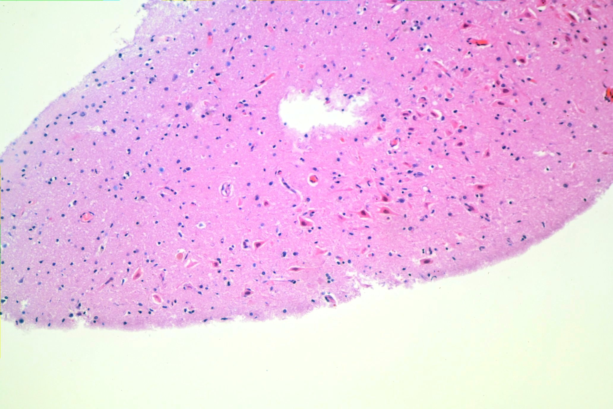
Here is a photomicrograph of neurons from the subiculum of a decedent who suffered a drug-related collapse with survival of a few days:
Here’s a close up:
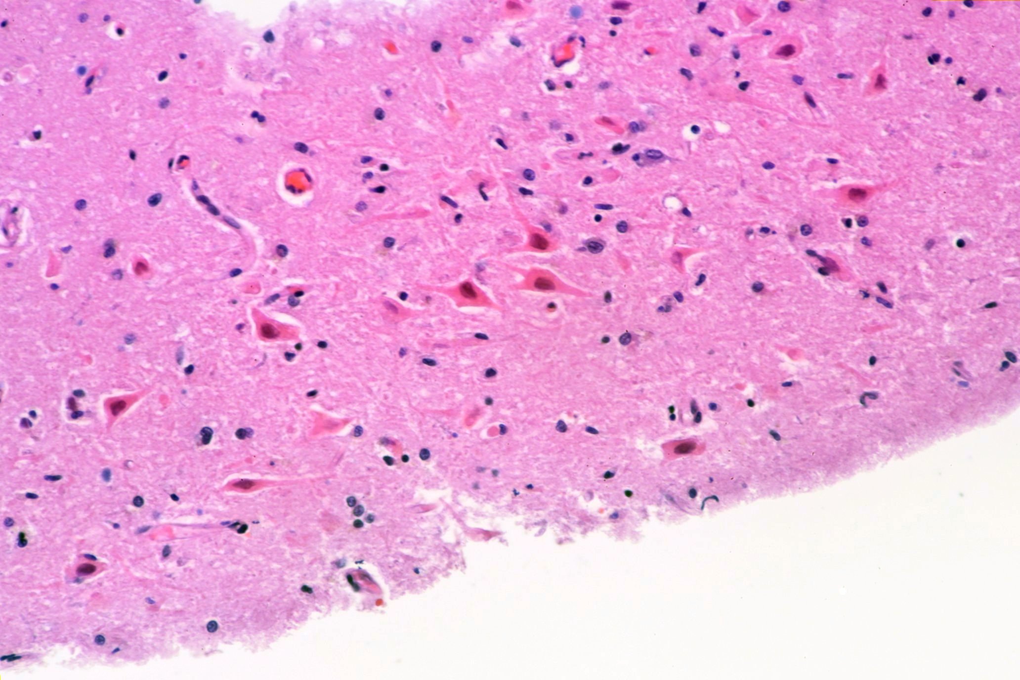
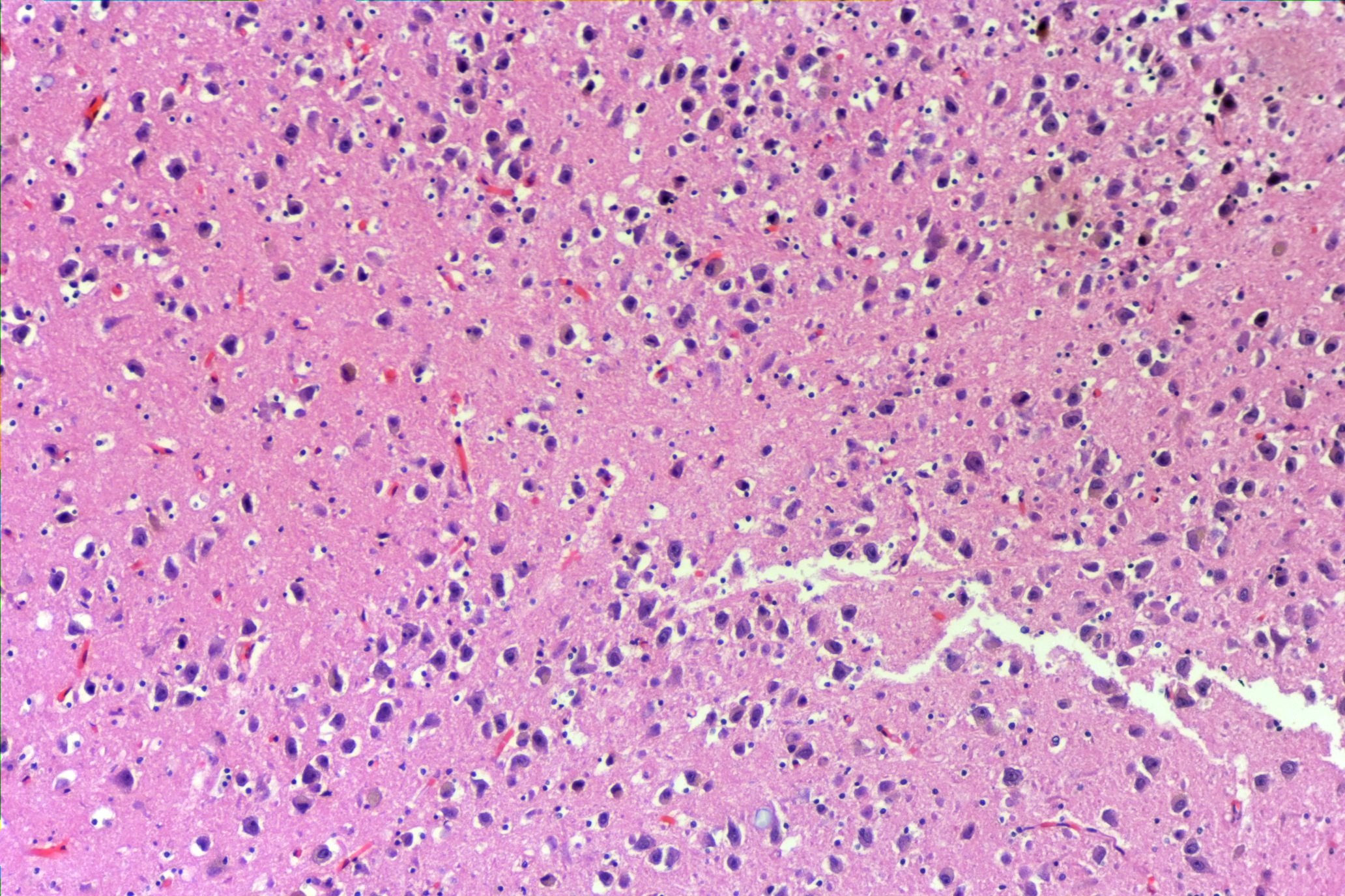
You can see how red, or “eosinophilic” the neurons are. Here’s a comparison from a person who died more suddenly:
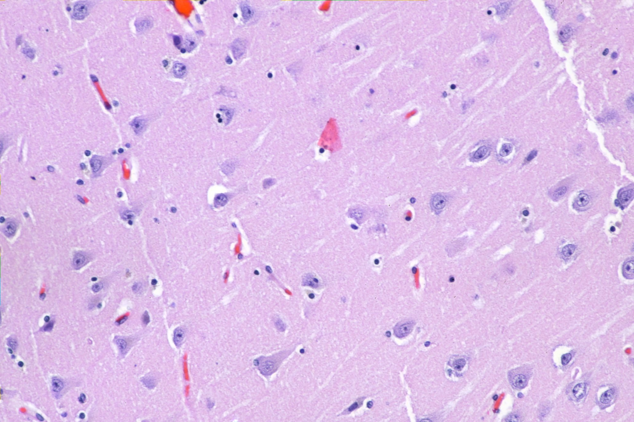
You can see how much more blue, or “basophilic” the neurons are.
In my experience, most drug deaths do *not* show red neurons. Instead there are different, and less specific changes such as darkening of the neuron and loss of nuclear definition and structure. But the textbooks love to talk about red neurons.