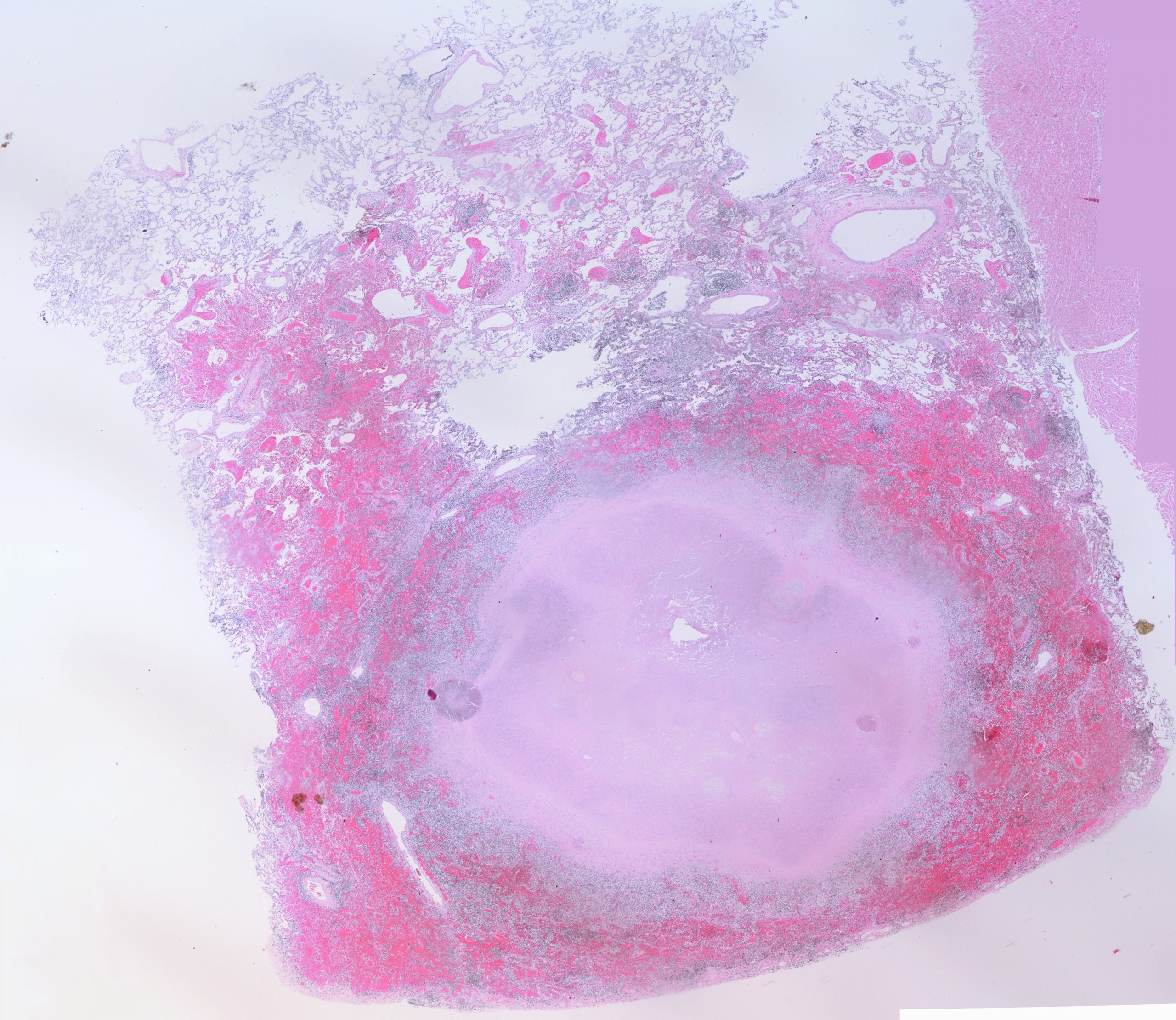
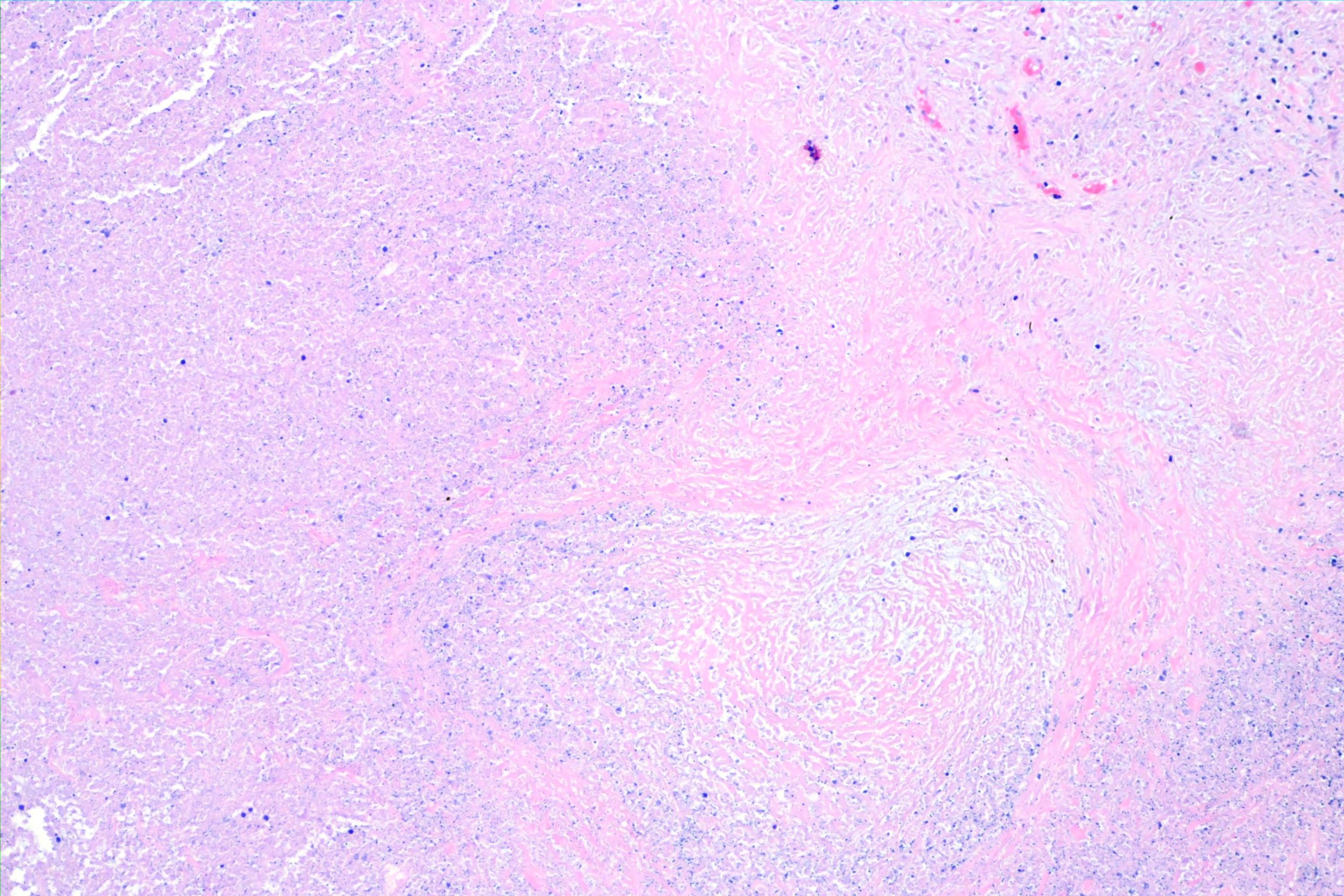
This is a pleural nodule found incidentally in a middle-aged man who died of a myocardial infarction. Here’s a panorama of the whole section. As always, click on the image to make it bigger (at least that’s what happens on my computer using Brave browser
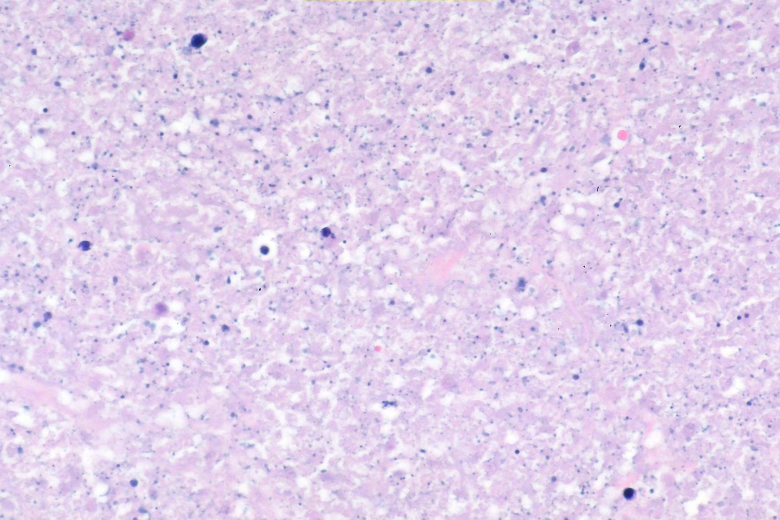
Here progressively higher power H&E stains:
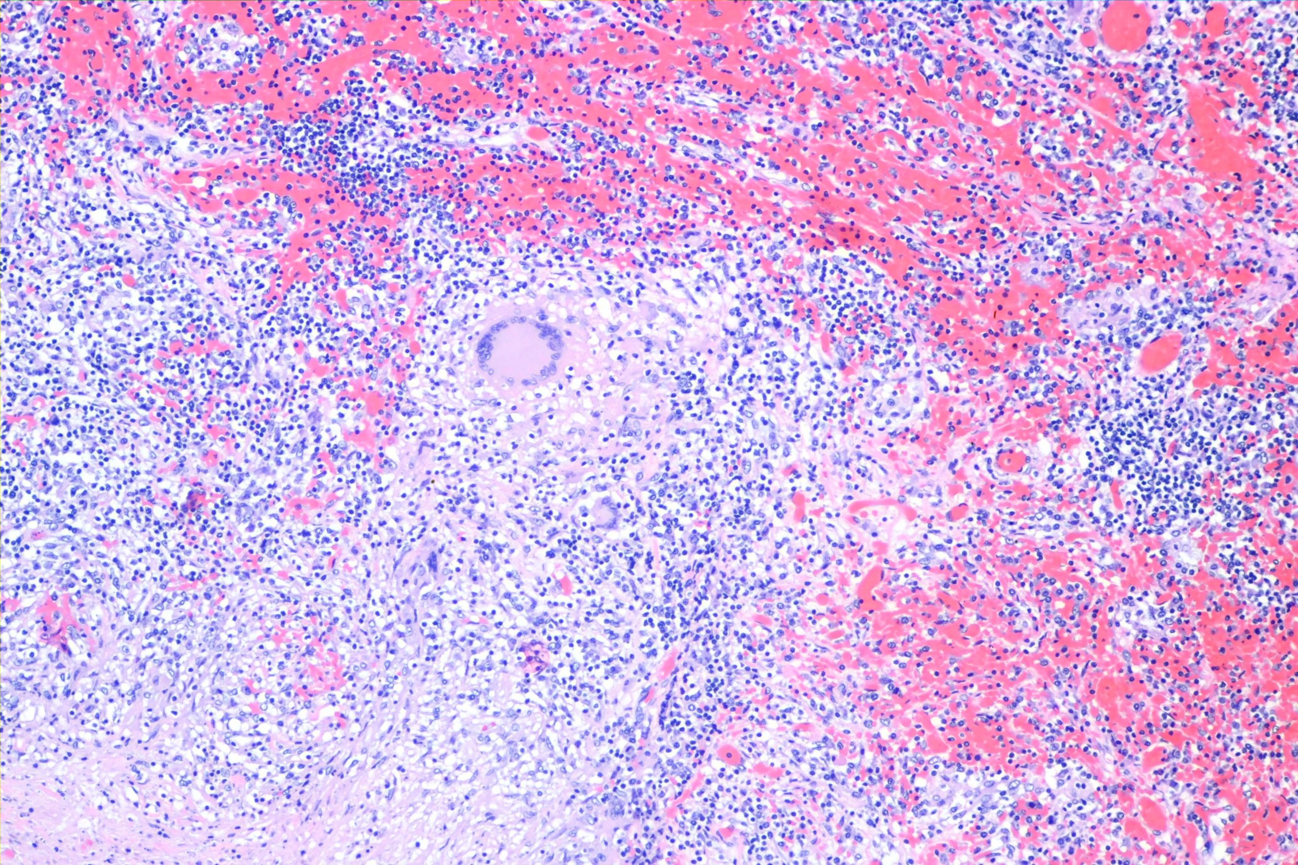
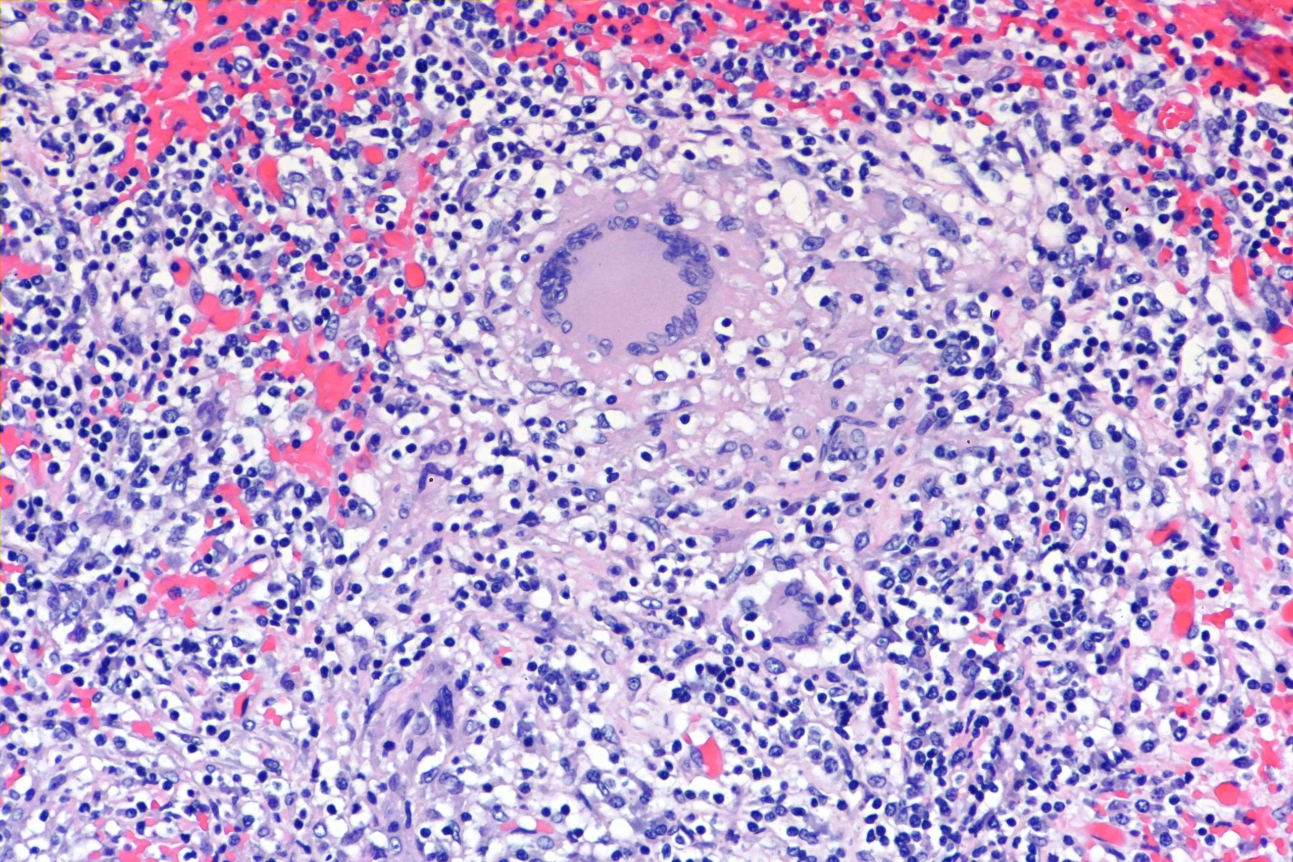
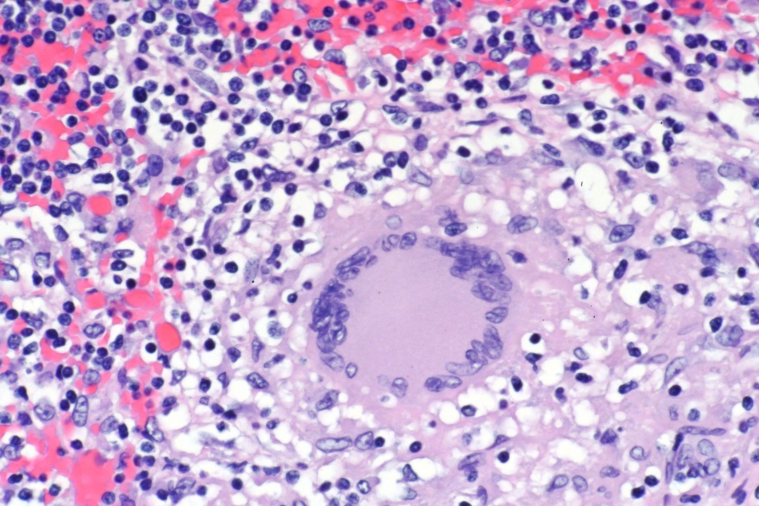
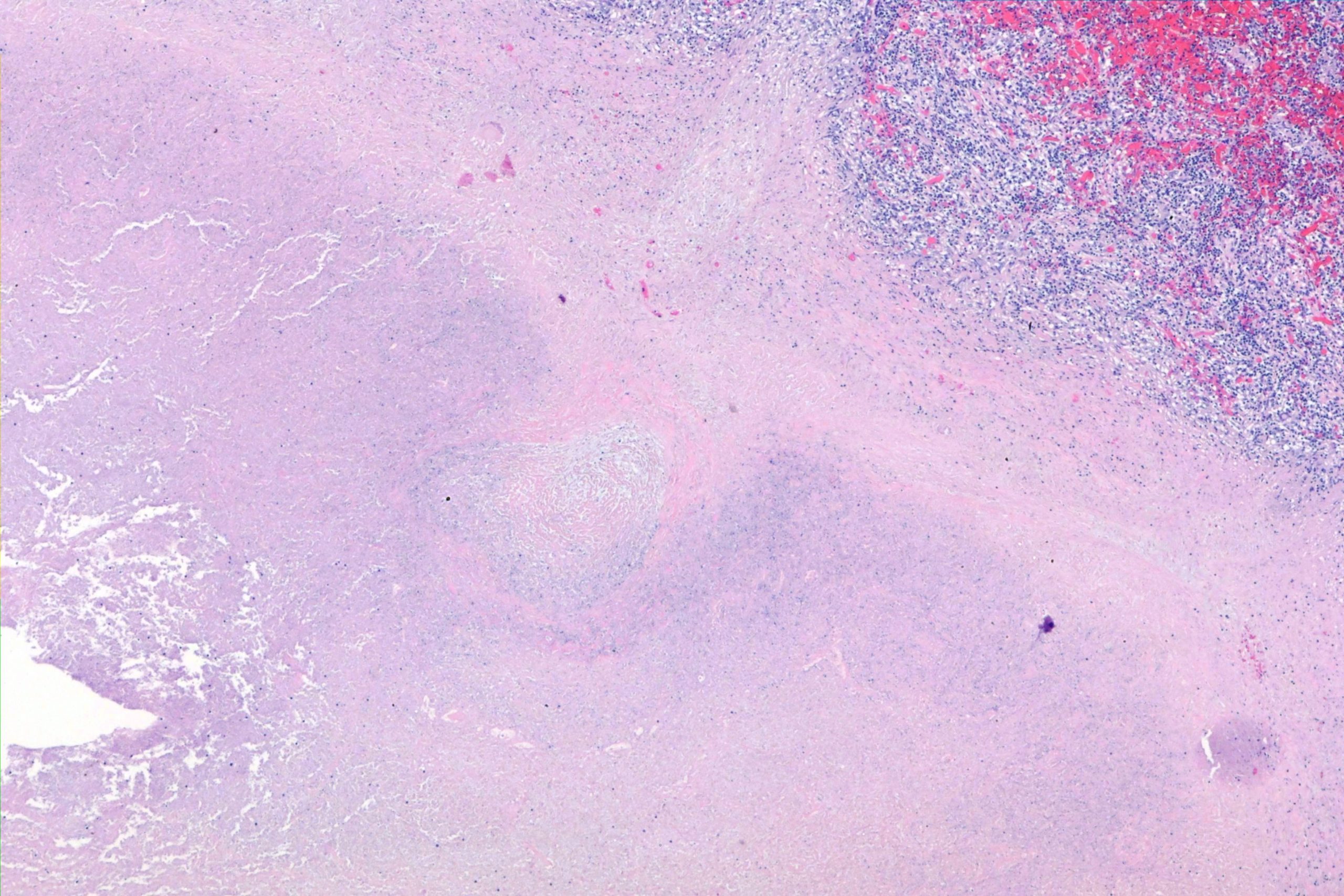
Here’s the area of caseous necrosis:
Here’s the obligatory GMS stain:
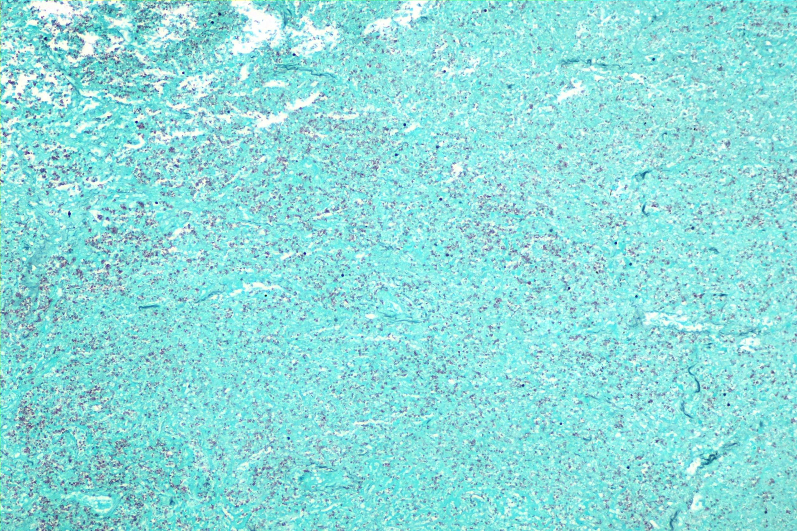
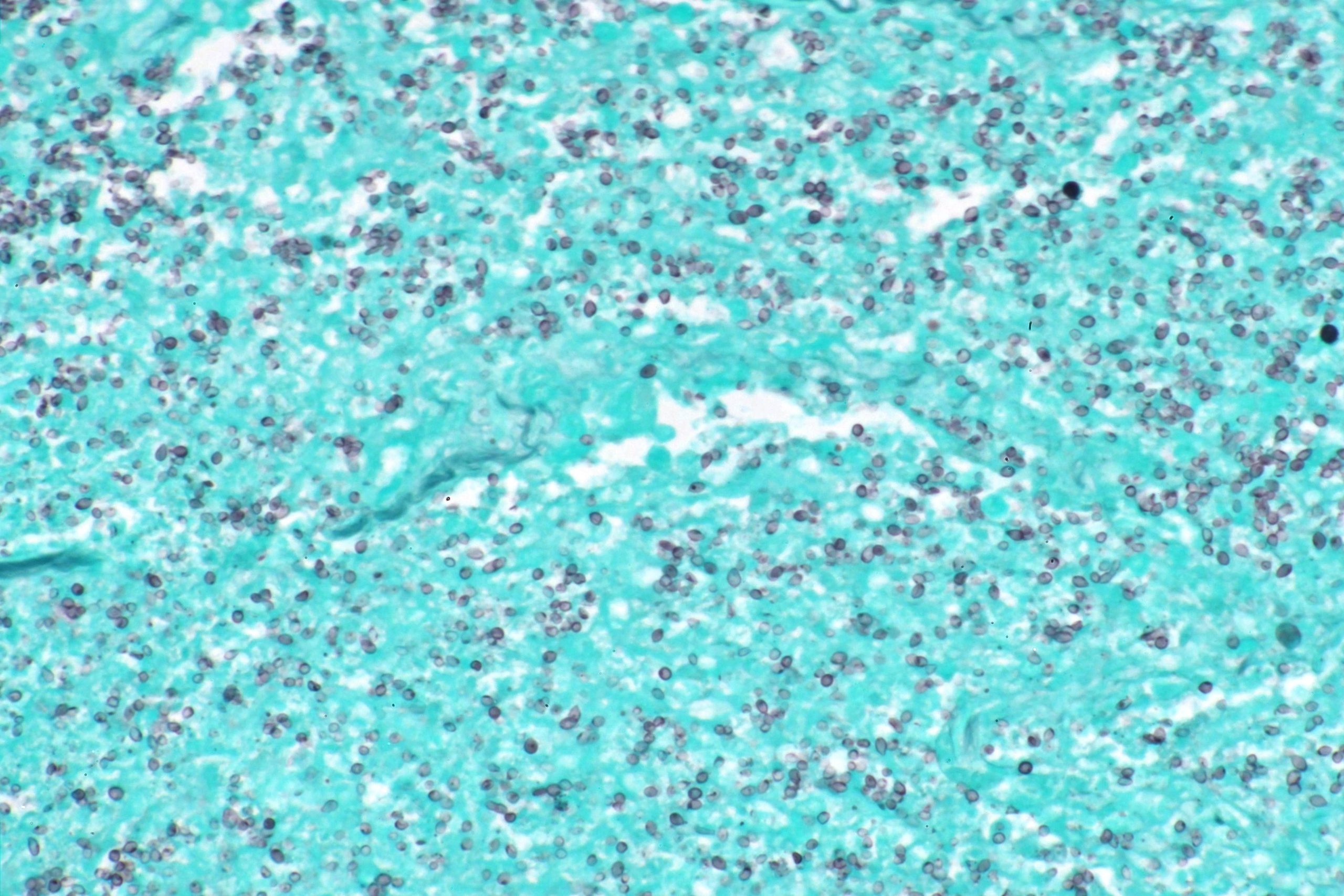
One thing that students and residents sometimes have a problem with is differentiating this from the gunk that stains black all the time — anthracosis, debris, etc. Students read that Histoplasma is very tiny, so every little dot looks like a yeast form. But it’s not. As you can see, they *are* pretty small, but even so they demonstrate the characteristic cell wall appearance and occasional unambiguous budding form.