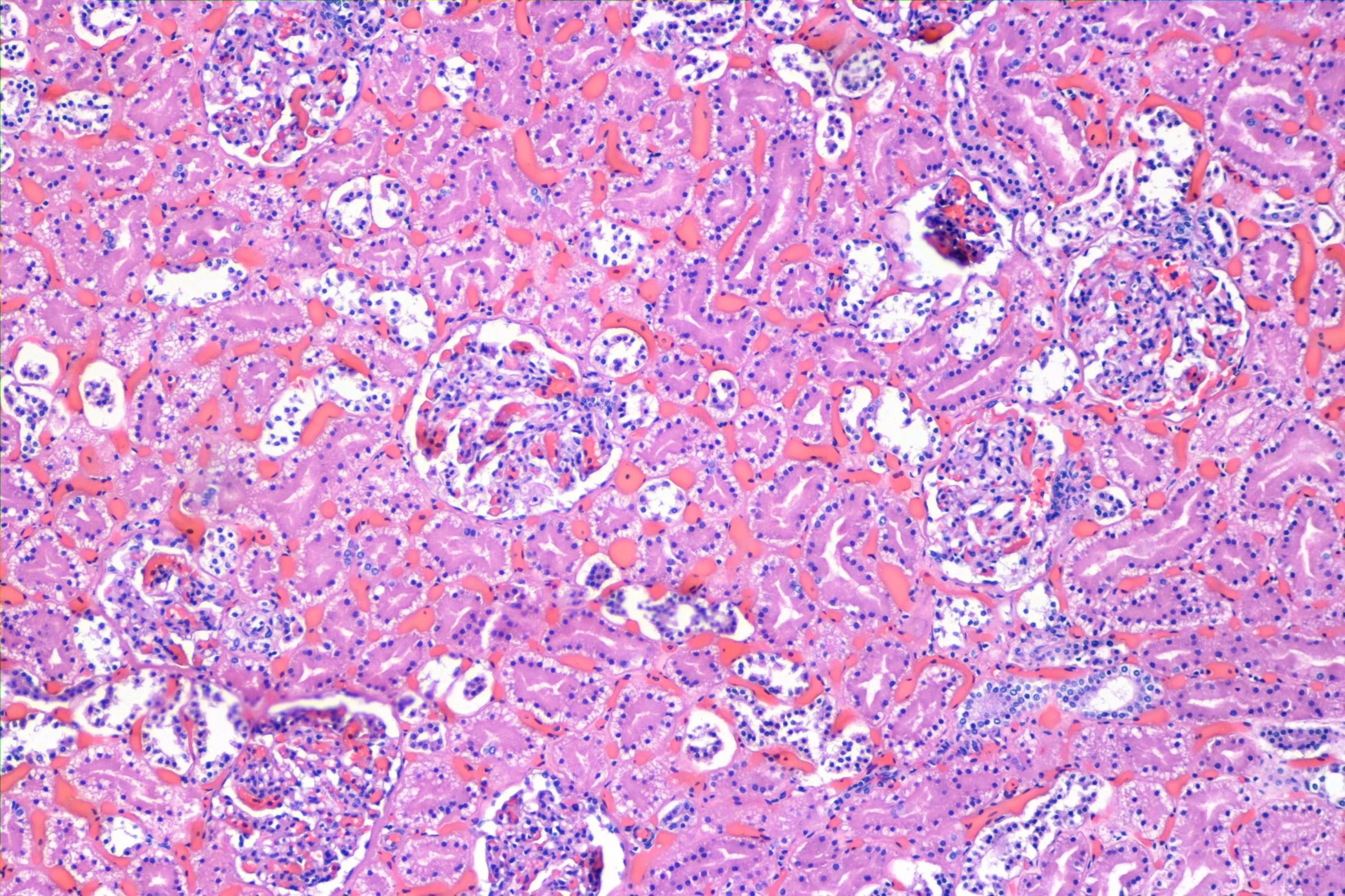
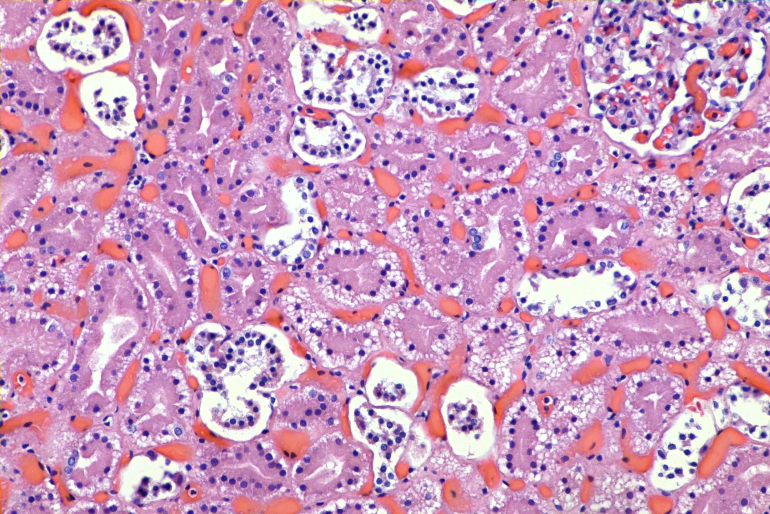
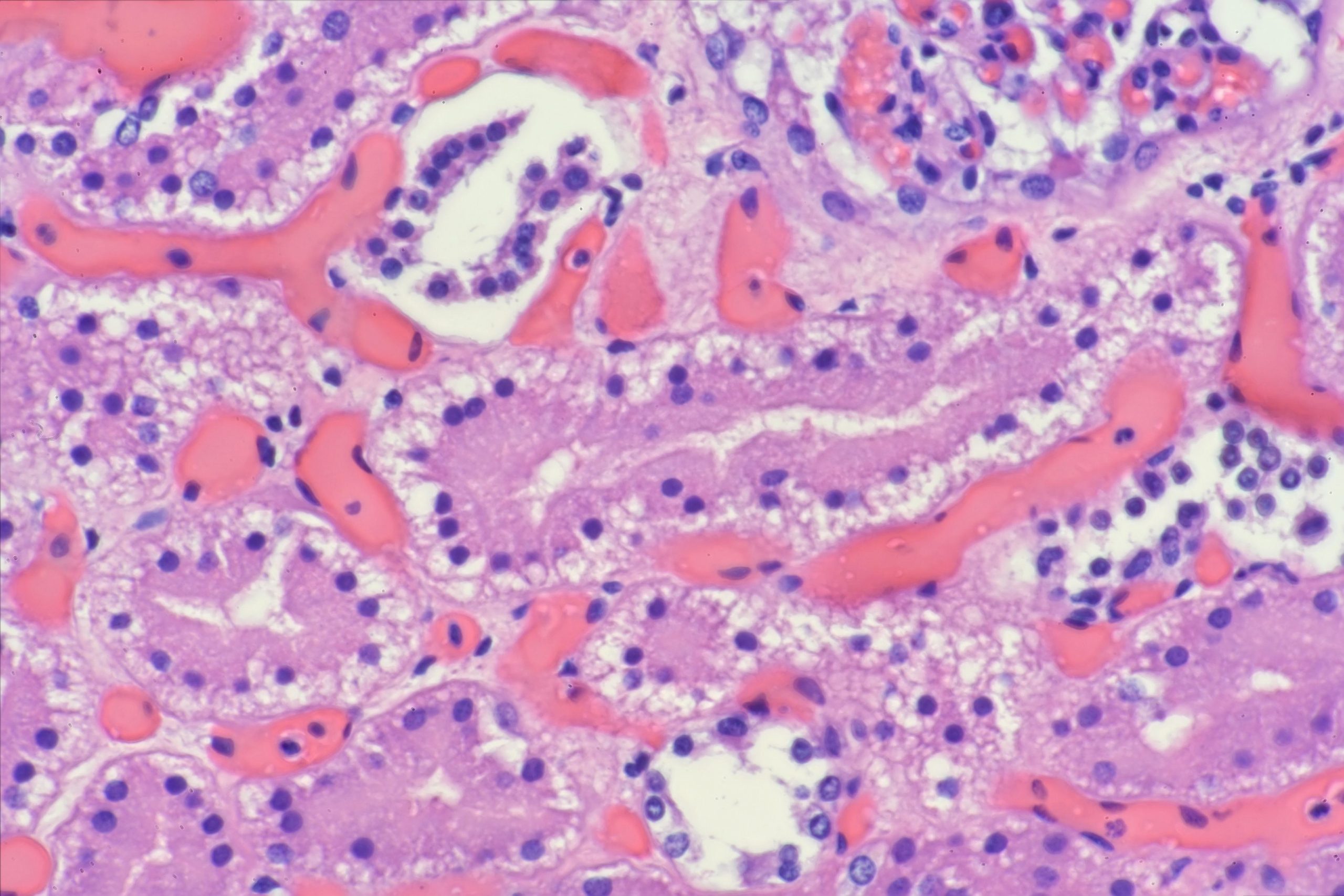
Nice example of basilar vacuolization of the proximal tubules of the kidney in a case of diabetic ketoacidosis. This is commonly called an “Armanni-Ebstein” lesion, though the original article by Armanni and Ebstein seems to have described a different effect, that of increased glycogen, and were more dispersed. These contain lipid.

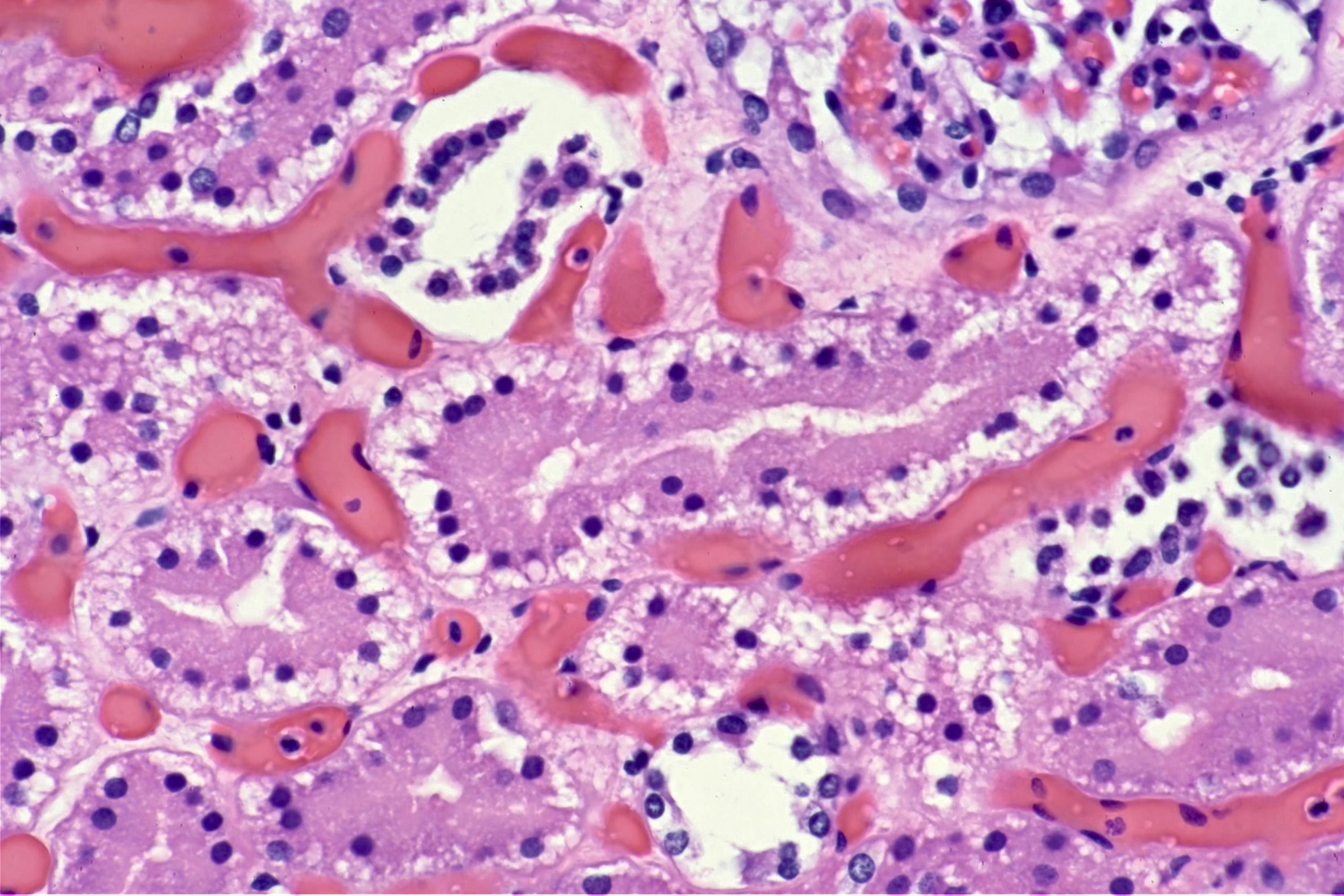
Just for fun, here is a stack-focused version of the above area. To really see the difference, look at the nuclei in the distal tubule at the right margin of the image, halfway down the side. This was done using the “Stack Focuser” plugin in ImageJ/Fiji.
Update:
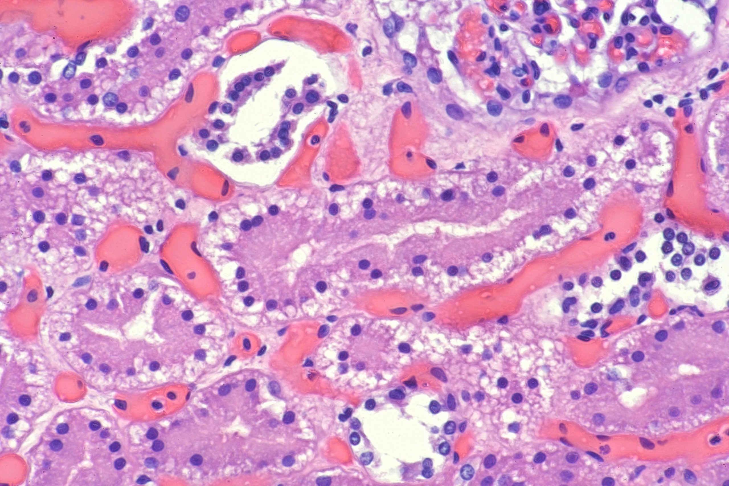
Here’s a focus stack using the same images, but with the “enfuse” program with high contrast weighting. While the Fiji plugin looks crisper, it also has hard edges, which are obvious if you click on the image and look at the full image. The enfuse version smooths those edges.
As always, free for use in lecture, or teaching, with or without attribution (though attribution is appreciated). If you put these in a publication, please contact me. Higher resolution images with lossless compression are available on request until I lose them, if you need them for a lecture or such. Email me if you want me to send them to you.
Billo,
Absolutely phenomenal!! Beautiful presentation!
Pete
I agree with Pete