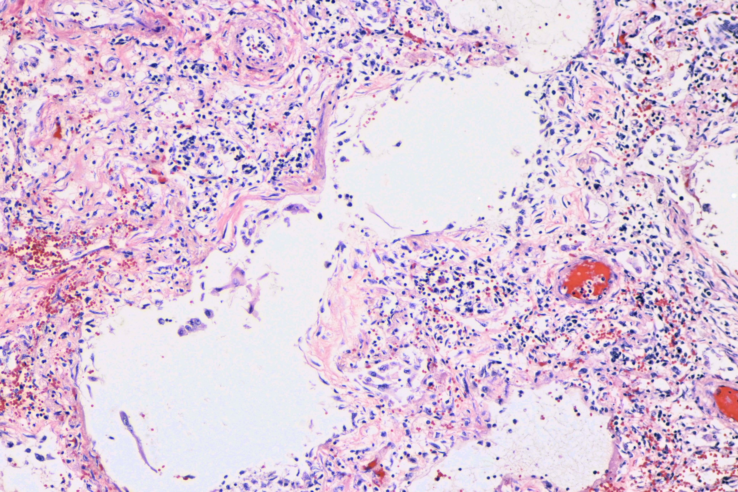
Here are some photomicrographs of the lung of a 65-year-old man who came into the hospital short of breath. He tested SARS-Cov-2 positive. His condition deteriorated, complicated by deep vein thrombosis, pulmonary thromboemboli, and cerebrovascular accident. At autopsy, he had, among other things, characteristic lung findings of diffuse alveolar damage with some organization as well as scattered fibrin microthrombi.
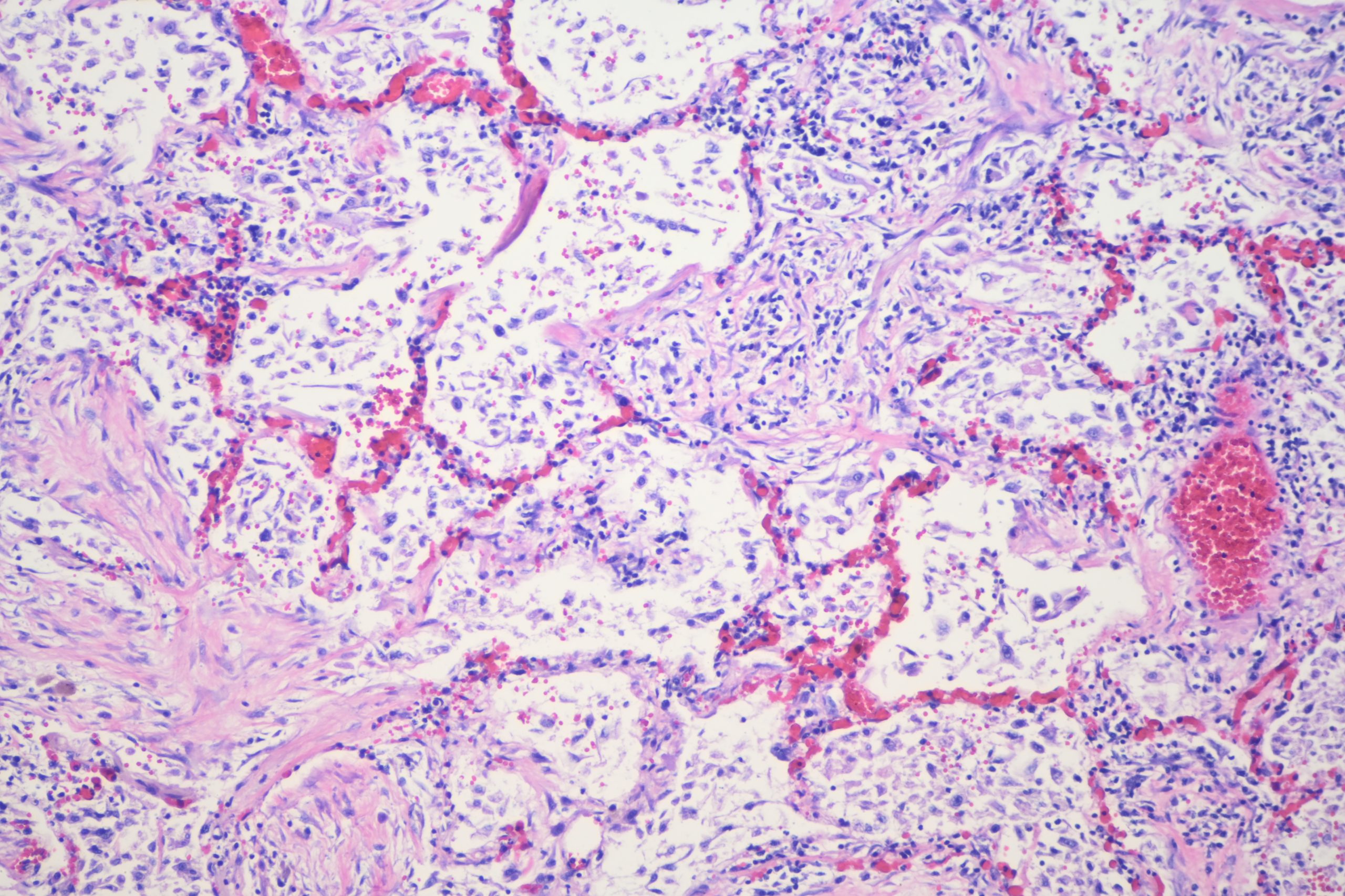
Here are some photomics of the lung.
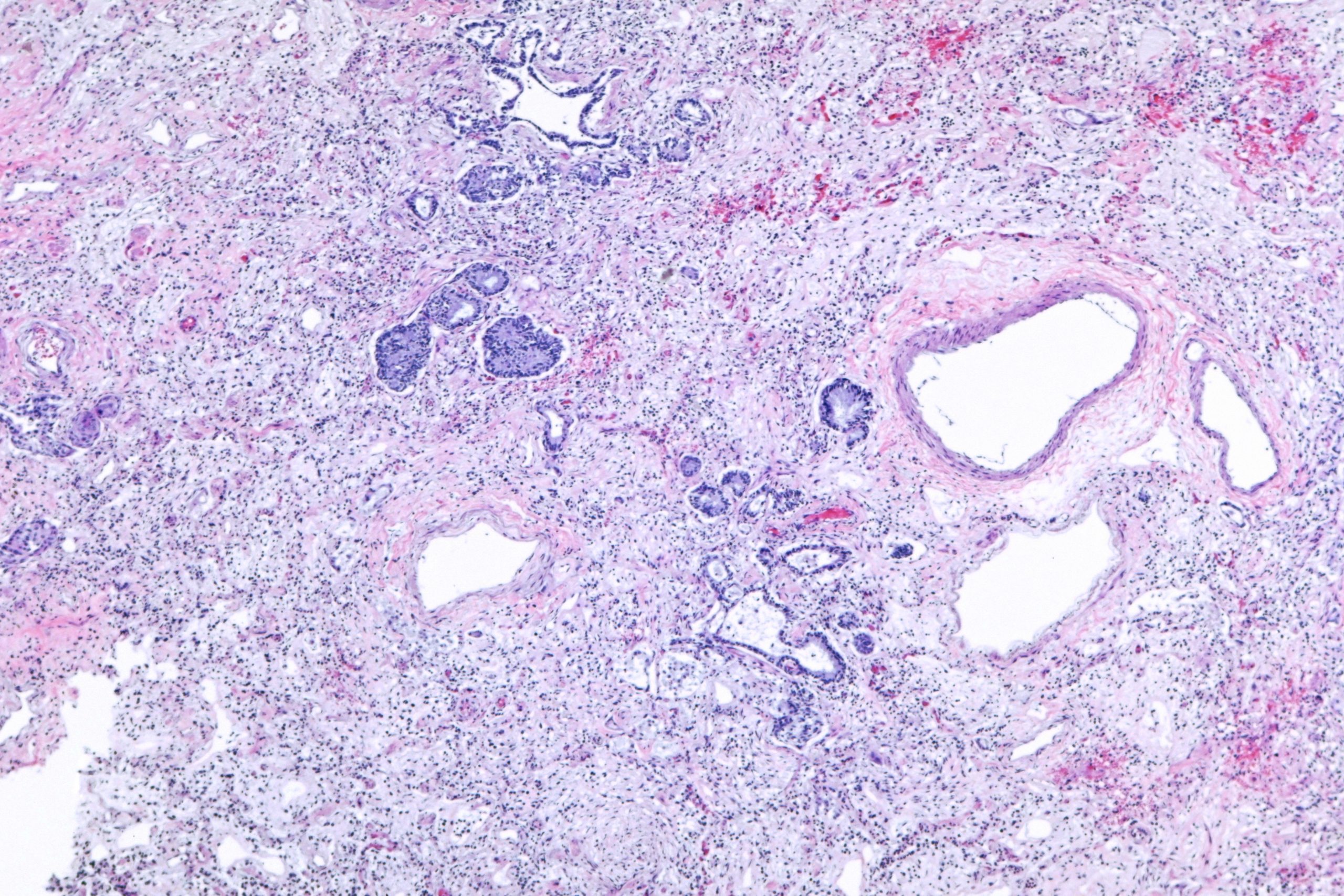
Low power mosaic:

binary comment
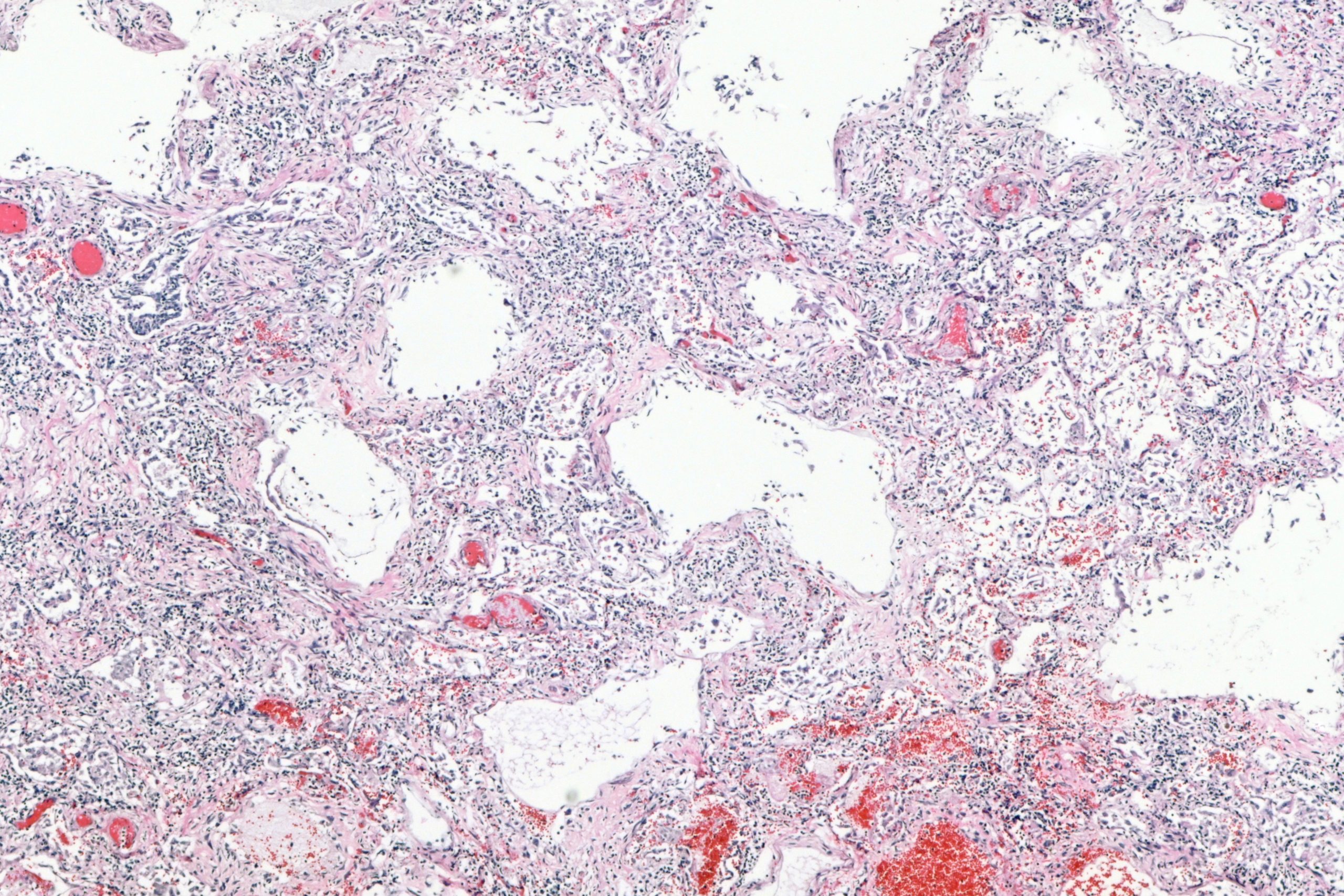
Some closer views of the alevoli, with fibrosis.

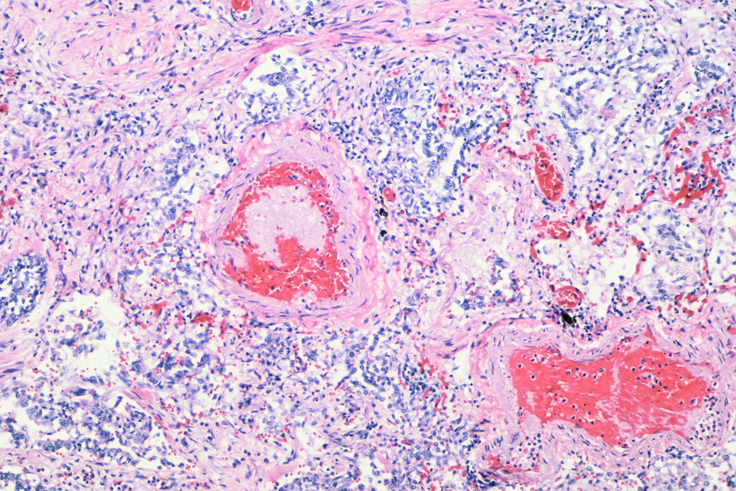
Some fibrin in a small vessel:
A few more views:
As always, free for use in lecture, or teaching, with or without attribution (though attribution is appreciated). If you put these in a publication, please contact me. Higher resolution images with lossless compression are available on request until I lose them, if you need them for a lecture or such. In particular, the whole mount view is about 21Kx11K pixels in the original TIFF image. If you need that big of an image I will have to figure out how to get it to you. Email me if you want me to send them to you.
Billo,
Beautiful presentation!! Was that your own case?
Question: Would an anticoagulant such as Eliquis possibly have been appropriate treatment?
Pete
Nice case. Thank you for sharing.