This is a series of images that shows what one can expect to see in cases of aspiration of gastric contents. Aspiration of gastric contents is very common in forensic cases. Back in the 1960s, people used to hear that famous musicians overdosed on drugs and “choked on their own vomit.” This was not true, as regurgitation is a common event in death, and it is also a common result of resuscitative efforts. Nowadays, unless there’s some other indication that it really was involved in the death, it’s considered essentially an artifact of death.
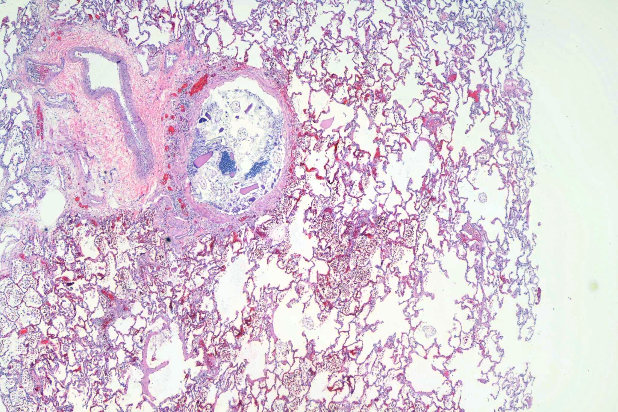
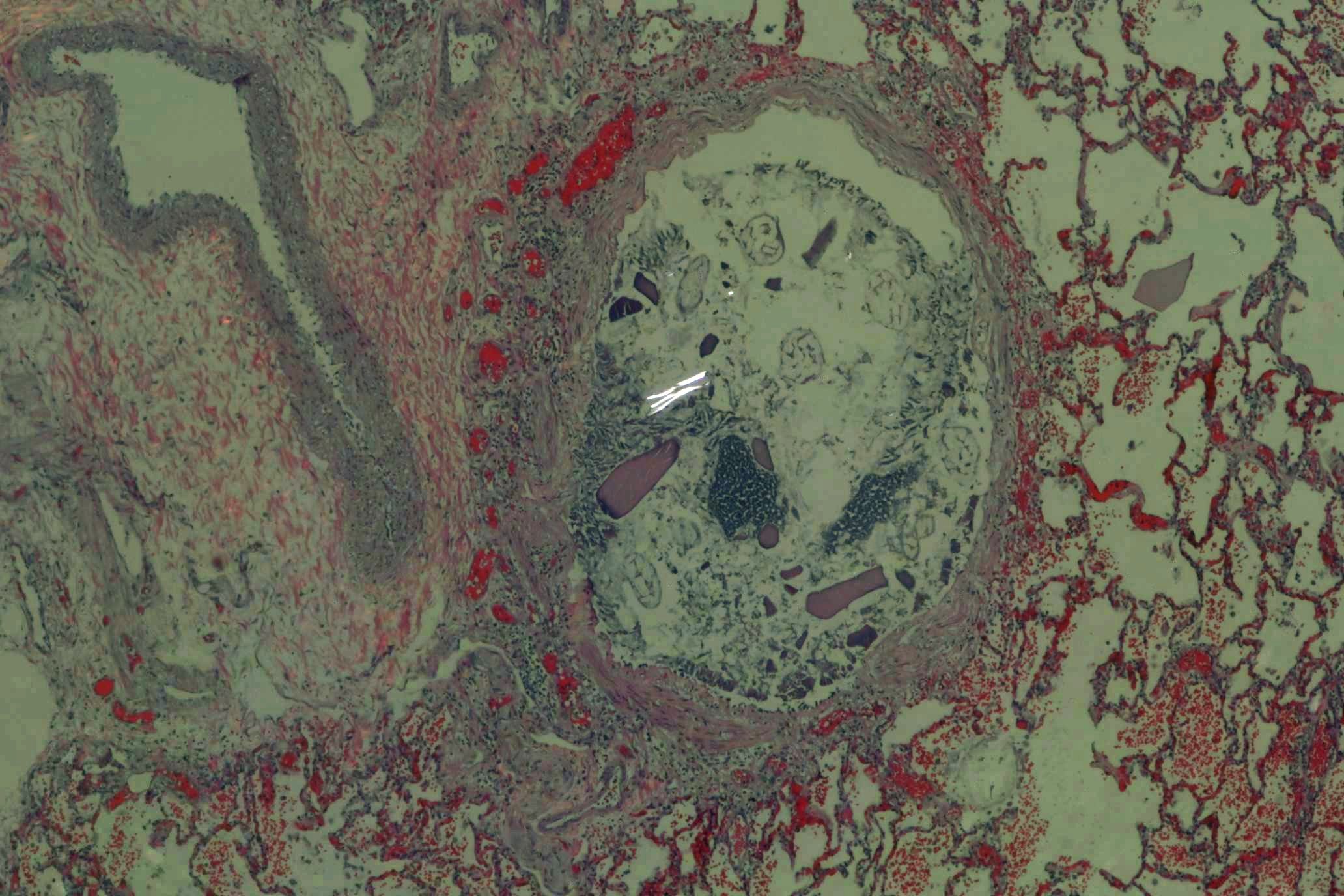
Here is a relatively low power view of a rather large bronchiole (no cartilage) filled with stomach contents from a fentanyl death:
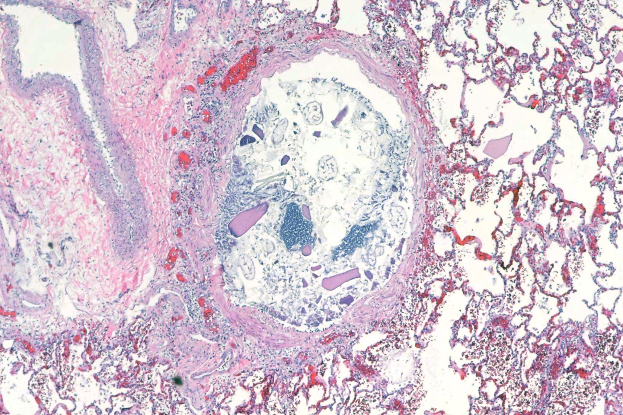
Here’s are successively higher power views:
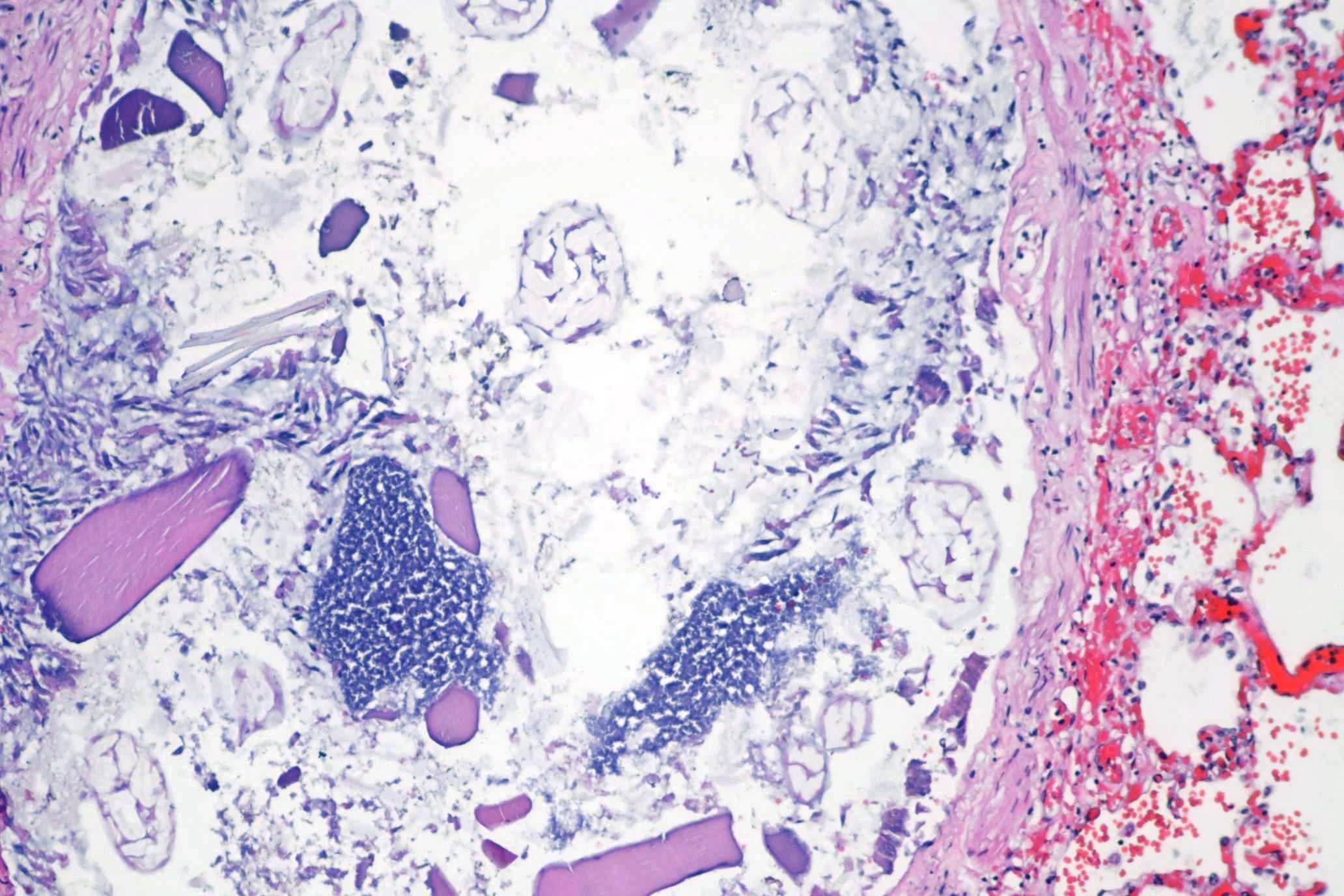
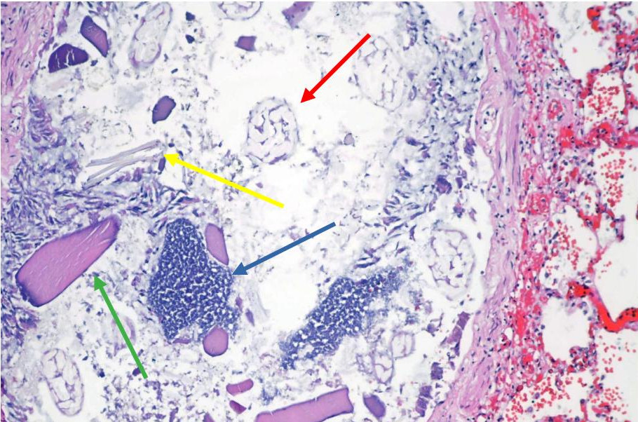
The material is easily identified. In the annotated image below:
- Plant material with easily identifiable cell walls (red arrow)
- Striated muscle (green arrow)
- Bacterial clusters (blue arrow)
- Amorphous debris (yellow arrow)
The amorphous debris is often not organic, and can be birefringent:
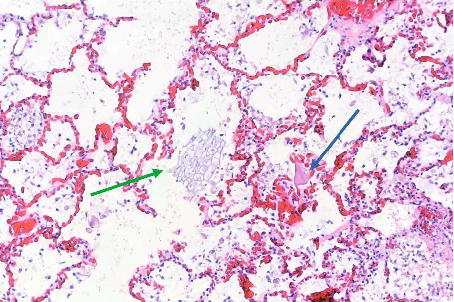
Gastric contents are often found outside of the larger airways, extending into alveoli. Here’s an example of plant material (green arrow) and meat (blue arrow) in alveolar spaces.
As always, free for use in lectures, etc. Attribution is appreciated but not required. The images are taken at high resolution (8Kx5K pixels), but reduced for the web. If you need high resolution images, contact me.
Nice photos, Billo. Steve Cohle