Here is a case of a young man who collapsed after sprinting. At autopsy, he was noted to have numerous sickled cells, sometimes clumping and filling vessels. Further workup revealed sickle trait.
Because the gene that makes sickle cell hemoglobin (Hemoglobin S) is autosomal, there are two copies in each cell. If both copies have the HgbS mutation, then the person has “sickle cell disease.” If the person has one normal and one HgbS, he or she has “sickle cell trait.” Sickle cell disease is life-threatening, painful, and debilitating. Sickle trait is largely asymptomatic. In fact, until the 1980s, it was believed that sickle trait was completely benign. Then folk noticed that people with sickle trait tended to collapse and die under very specific circumstances — when they exercise past the point of exhaustion. Thus, there was greatly increased mortality among people with sickle trait going through military training, athletic training, and when running from the police.
The problem is that sickle cells have a roughly normal shape when there’s lots of oxygen in the blood. When oxygen gets low, the cells collapse into shapes that easily clump, resulting in blockage of blood vessels and damage to tissues downstream of the blockage. Young cells exhibit “reversible” sickling — the collapse when oxygen starved, and expand into a more normal shape when oxygen is replenished. Older sickle cells are “irreversibly” sickled, and will not expand when exposed to oxygen.
For a more complete description, I discuss it a bit more in my evaluation of the George Floyd case .
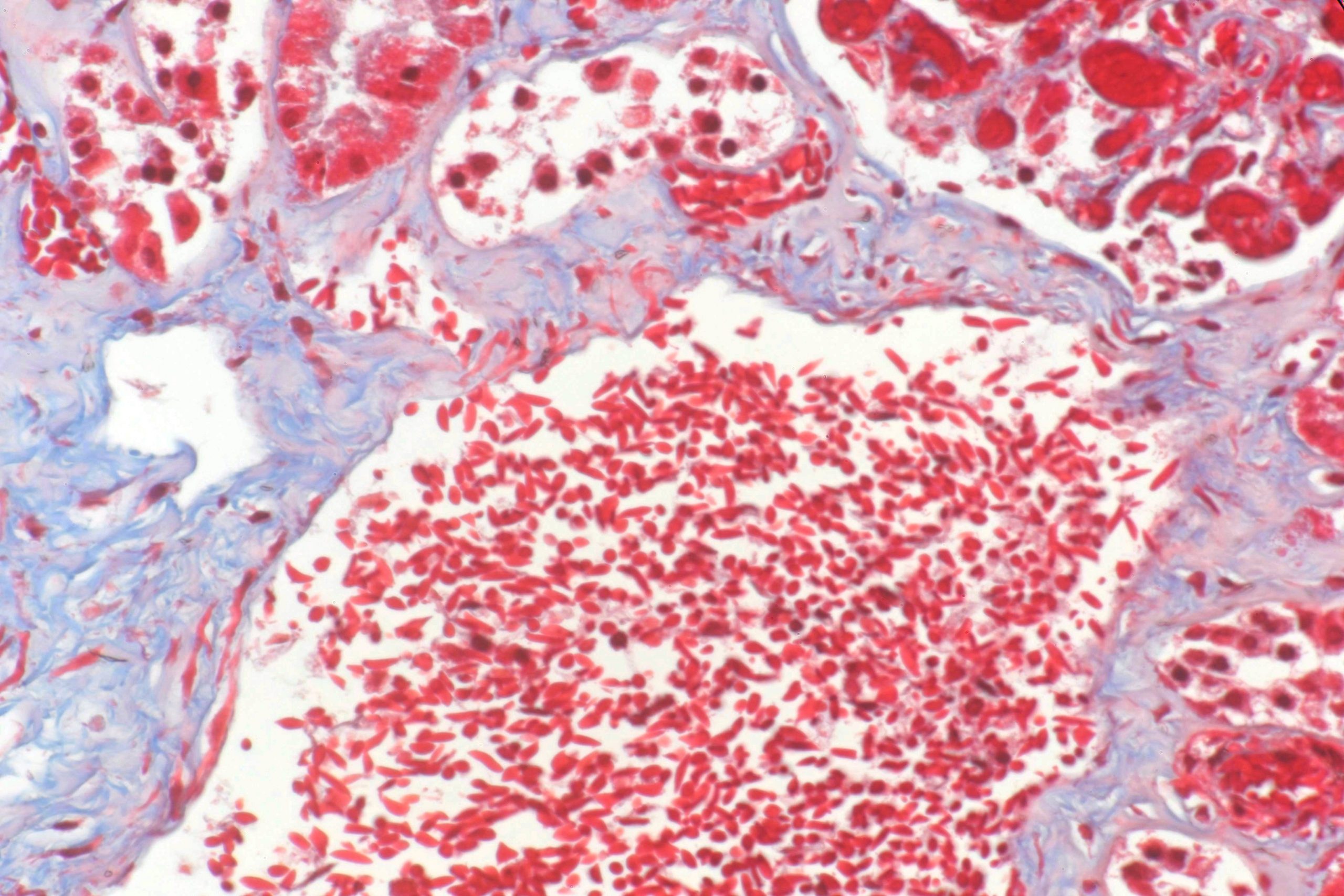
In this case, the decedent was a young man who collapsed after exertion and went into multiorgan failure — a common presentation. At autopsy, he demonstrated minimal pathology, but had abundant sickle cells in many organs. Here’s an example from the kidney.
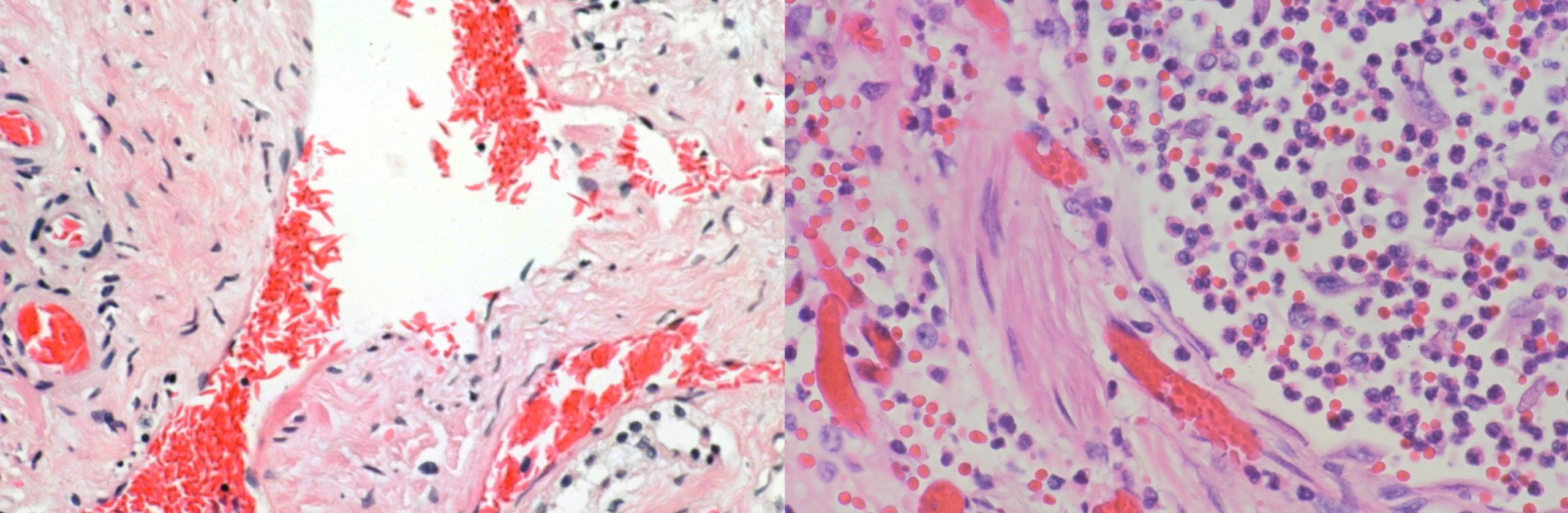
Here’s a side-by-side. On the left is a section from the decedent’s kidney showing thin, spiked red cells. On the right is a section from a decedent without sickle trait who died of pneumonia. Notice how round and plump the red cells are on the right. This is a large image and is scaled down automatically by WordPress for this web page. Right click on the image and open it in a new tab to see higher resolution.
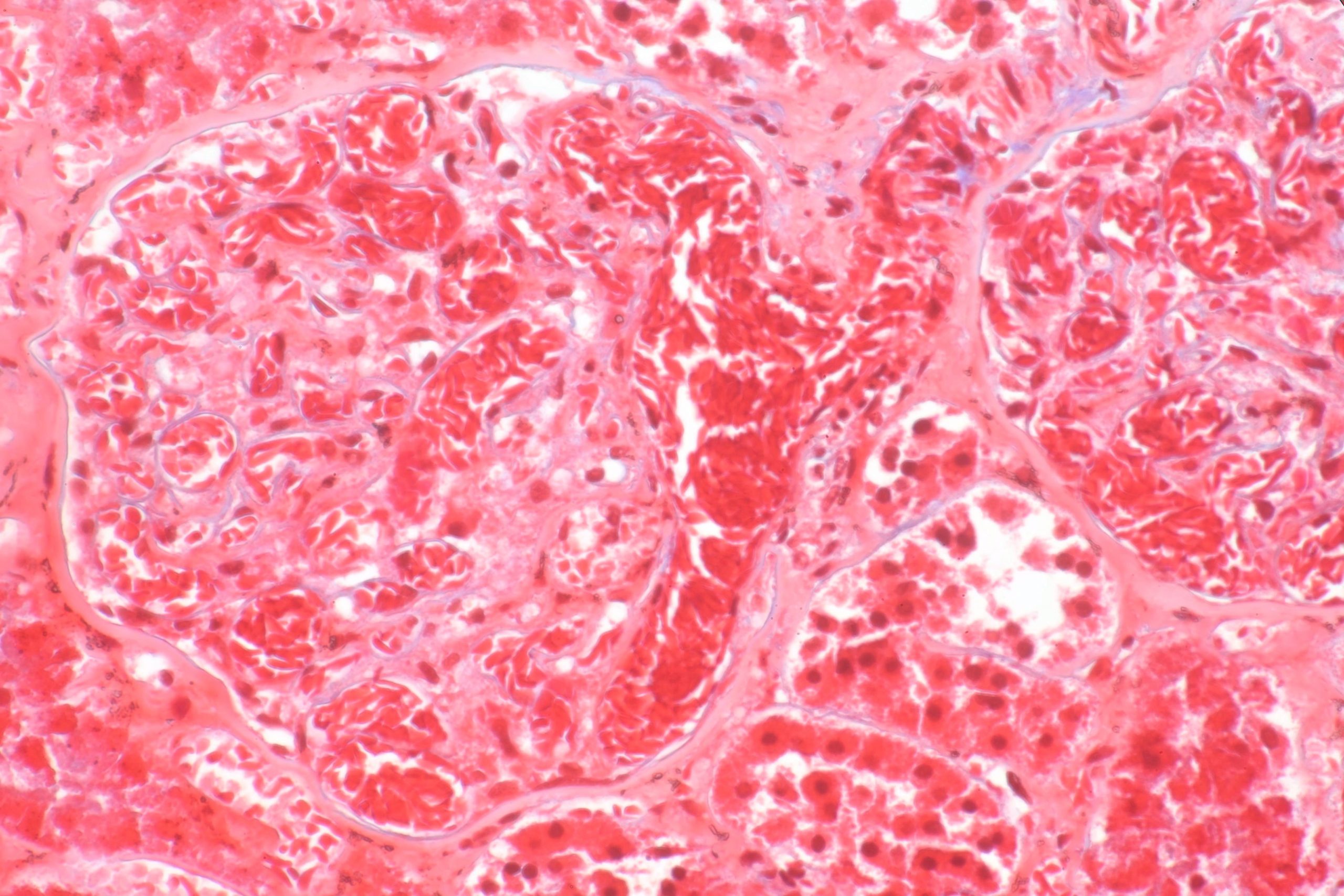
Here’s a picture of a renal glomerulus. You can see the sickled cells stacking up and blocking vessels. This is a trichrome stain.
One of the conundrums in postmortem evaluation of these cases is interpreting the presence of sickled cells in tissues. Some reports say that it’s not meaningful because the cells sickle in low oxygen environments, such as being dead. Thus, the presence of sickled cells does not itself imply a sickle crisis. On the other hand, other studies indicate that sickled cells can reverse upon tissue processing or drawing blood because the cells are often exposed to air in processing, and thus reversibly sickled cells can plump up. Thus, the absence of sickled cells does not rule it out. The diagnosis of exertional sickle trait death in the absence of obvious ischemic changes in affected organs is based primarily on history in the context of a diagnosis of sickle trait.
As always, free for use with or without attribution, though attribution is appreciated. If you need higher resolution copies, contact me.