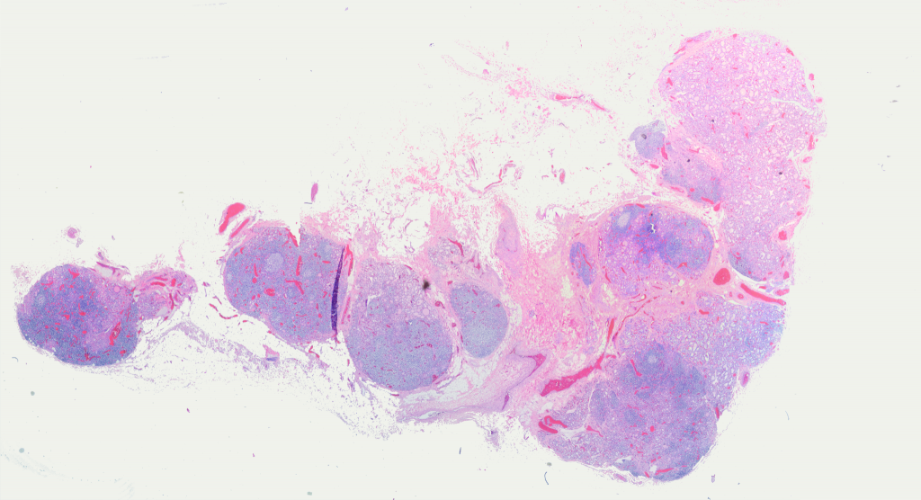
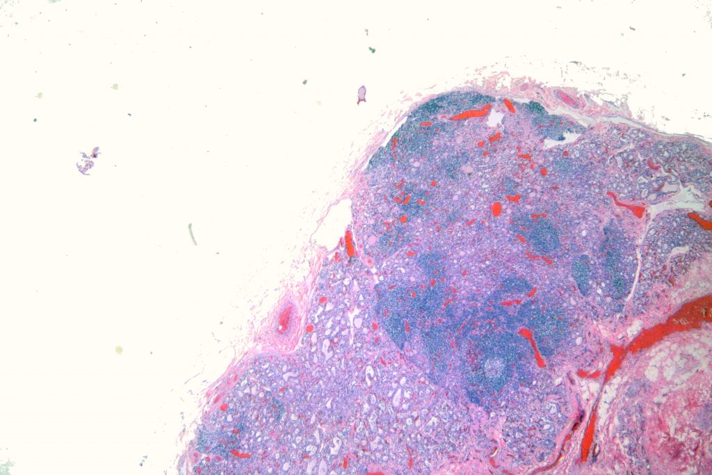
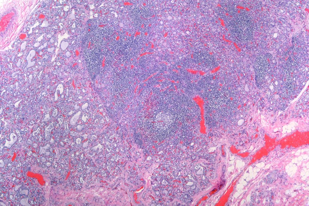
Here’s a classic example of severe lymphocytic thyroiditis in a middle-aged woman who died of unrelated causes. You can see in the whole slide panorama that much of the parenchyma of the thyroid has been replaced by lymphocytic aggregates with scattered germinal centers. Another common finding are so-called “Hurthle cells,” which are plump cells with eosinophilic cytoplasm and large nucleoli. The cytoplasmic change is said to by due to an abundance of mitochondria. I didn’t see them in this case. The textbooks say that the thyroid is often enlarged, but my experience is that it is more often pale and small. Maybe I only see late cases at autopsy. It often feels very firm to palpation if there’s some fibrosis.
Here’s the panorama shot:
Here’s a medium power:
And higher power. Note that the remaining follicles are small and a bit involuted. The parenchyma is largely replaced by lymphocytes with a prominent germinal center. Fibrosis is not a prominent feature here.


