Here are a couple of simple photomics of lipid emboli in the lungs. This is from a case of a young adult woman who died of methamphetamine and fentanyl intoxication. She was provided resuscitative efforts for about 30 minutes, including a mechanical resuscitator. At autopsy, she had small fractures of ribs 3-6 anterolaterally on the left.
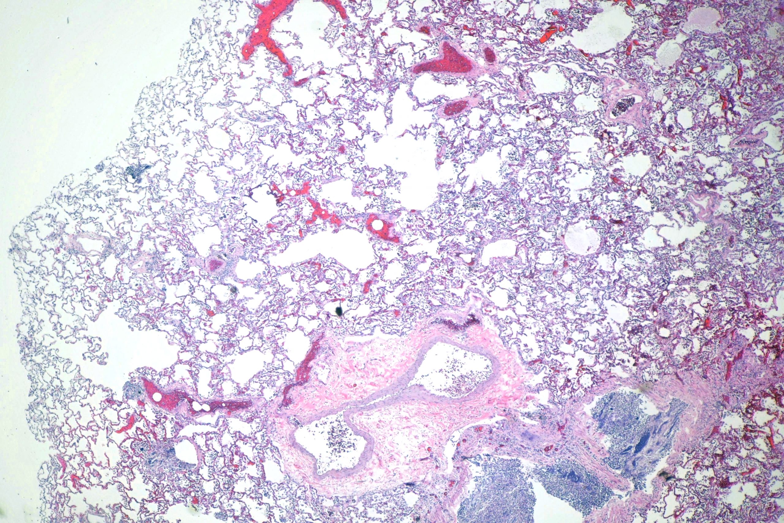
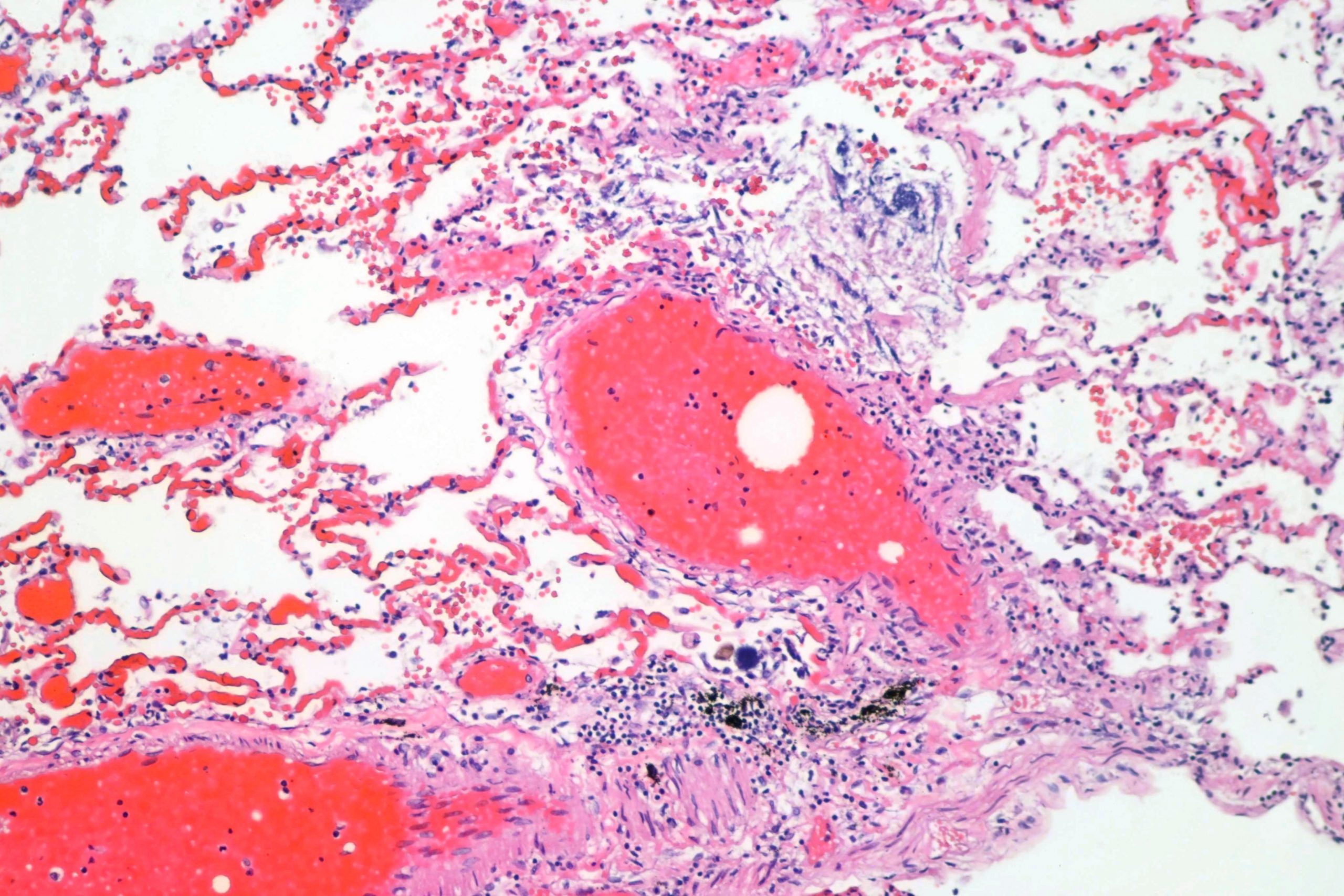
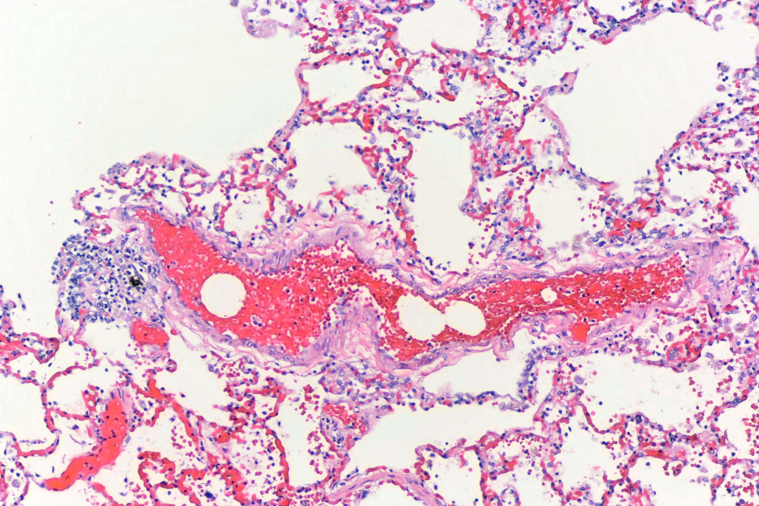
On histologic examination of the lungs. numerous small arteries contained small clear vacuoles. These are characteristic of small lipid droplets that are leaked into the blood when a bone breaks. Sometimes (but not this time), sizeable pieces of bone marrow are leaked into the blood and you can see hematopoietic elements in the lungs as well. When large bones are broken or there is massive blunt trauma, enough lipid can leak into the blood to cause respiratory distress or even death. In cases such as this, however, that’s not a problem.
Other things can look like this, of course. The most common is air embolism. I had a case some years ago where air was inadvertently injected into the blood by a nurse, and the lungs looked a bit like this. However, when there are obvious broken ribs and nothing to suggest air or other competing cause, it’s almost certainly lipid. Back in the day, I would always confirm the lipid with Oil Red O or osmium stain. But… nobody seems to offer osmium staining any more (it’s dangerous, apparently), and few places will do Oil Red O. Plus, cost is always a bigger factor now that it used to be. So, today, I just call it lipid and move on.
Here’s a low power view:
Here are a couple of higher power shots:
As always, free for use with or without attribution, though attribution is appreciated. If you need a high resoluton image, contact me.