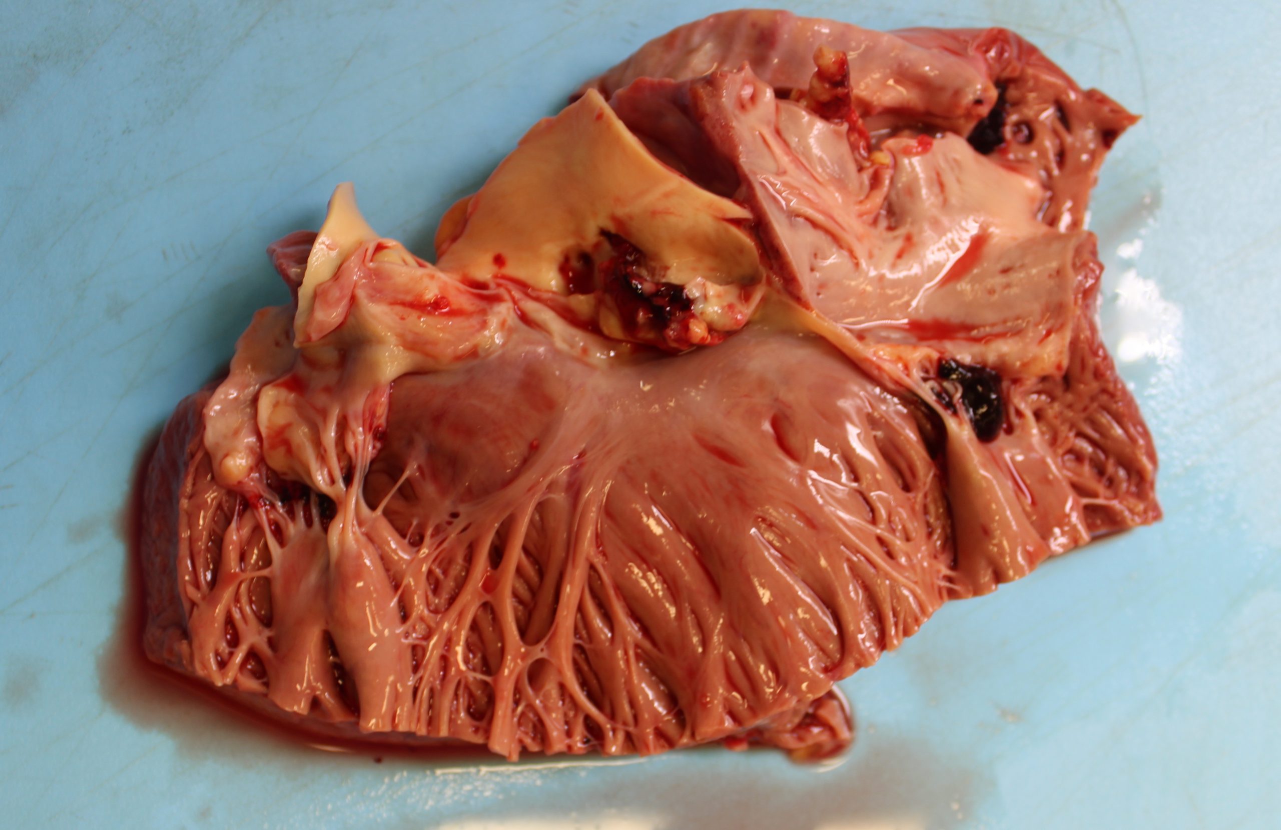
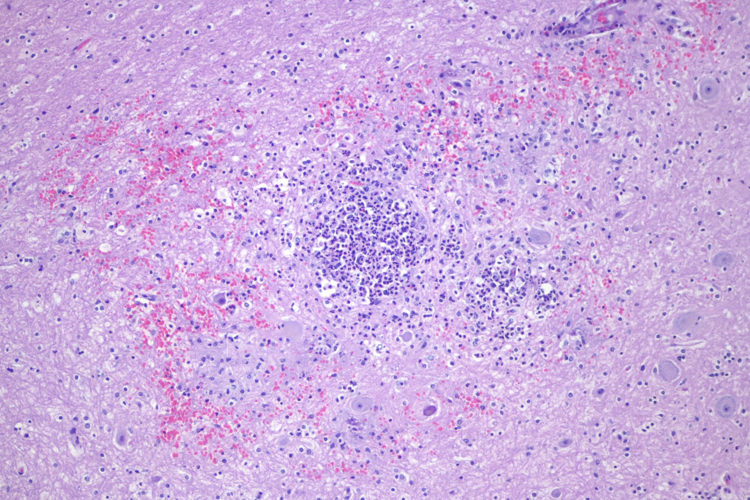
This is a repost from the now-defunct Pretty Pics series, with a couple of additions. I don’t remember the actual case any more, but the typical story is that of a young adult intravenous drug abuser with sepsis or some catastrophic failure (stroke, sudden arrhythmia, etc). At autopsy, there is a vegetation (or multiple vegetations) with microabscesses downstream. If the vegetation is on the right, then there’s typically a pneumonia, and if it’s on the left then multiple organs are seeded. In this case, there were microabscesses in multiple organs.
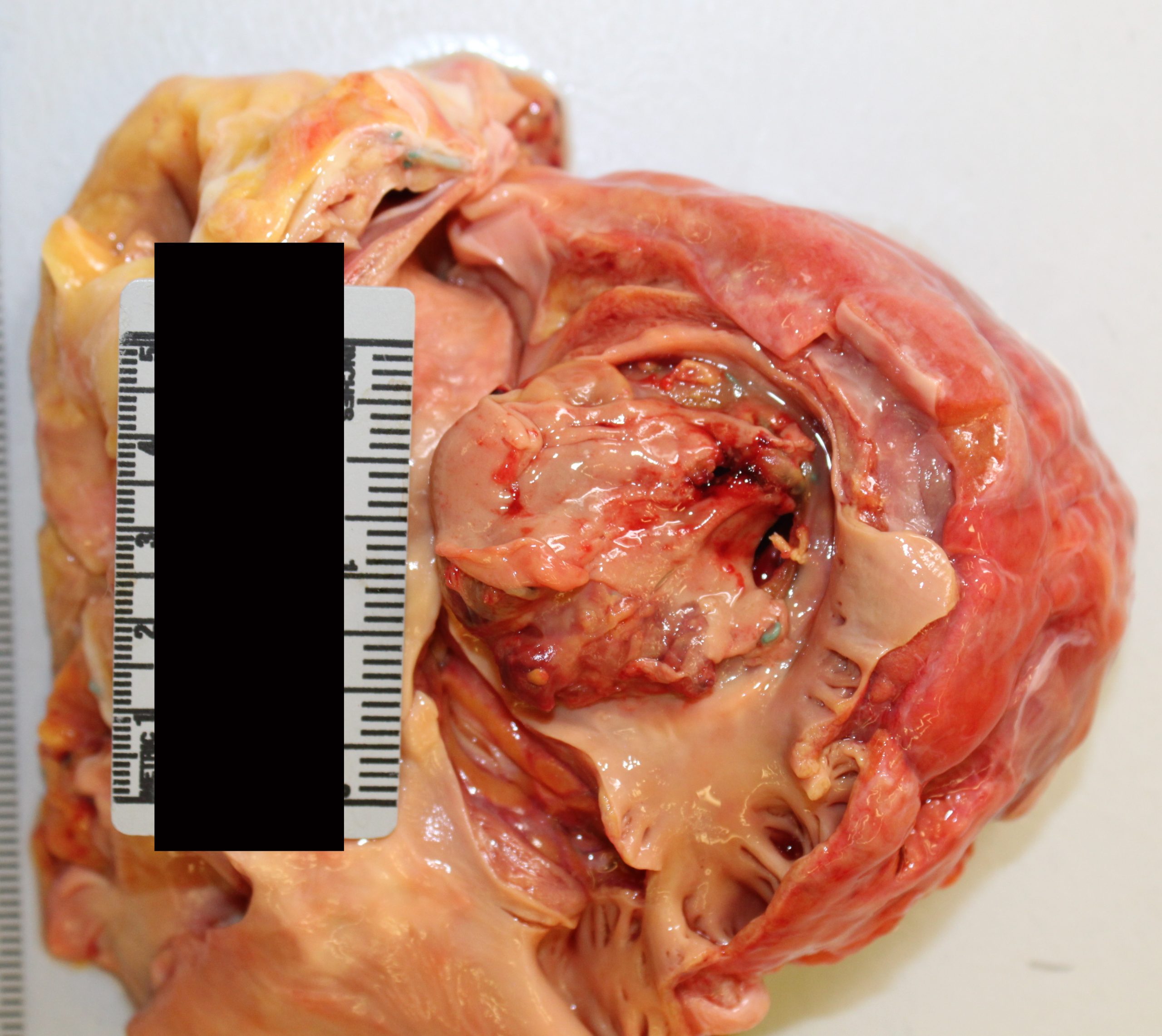
Grossly, these things look like they sound — sort of amorphous globs on a valve, with some adherent clot. I guess I’ve had about 30 or so of these cases in the past five years. Usually they aren’t all that big when I see them. Here are a couple of gross dissection pics of a couple of ones I’ve seen:
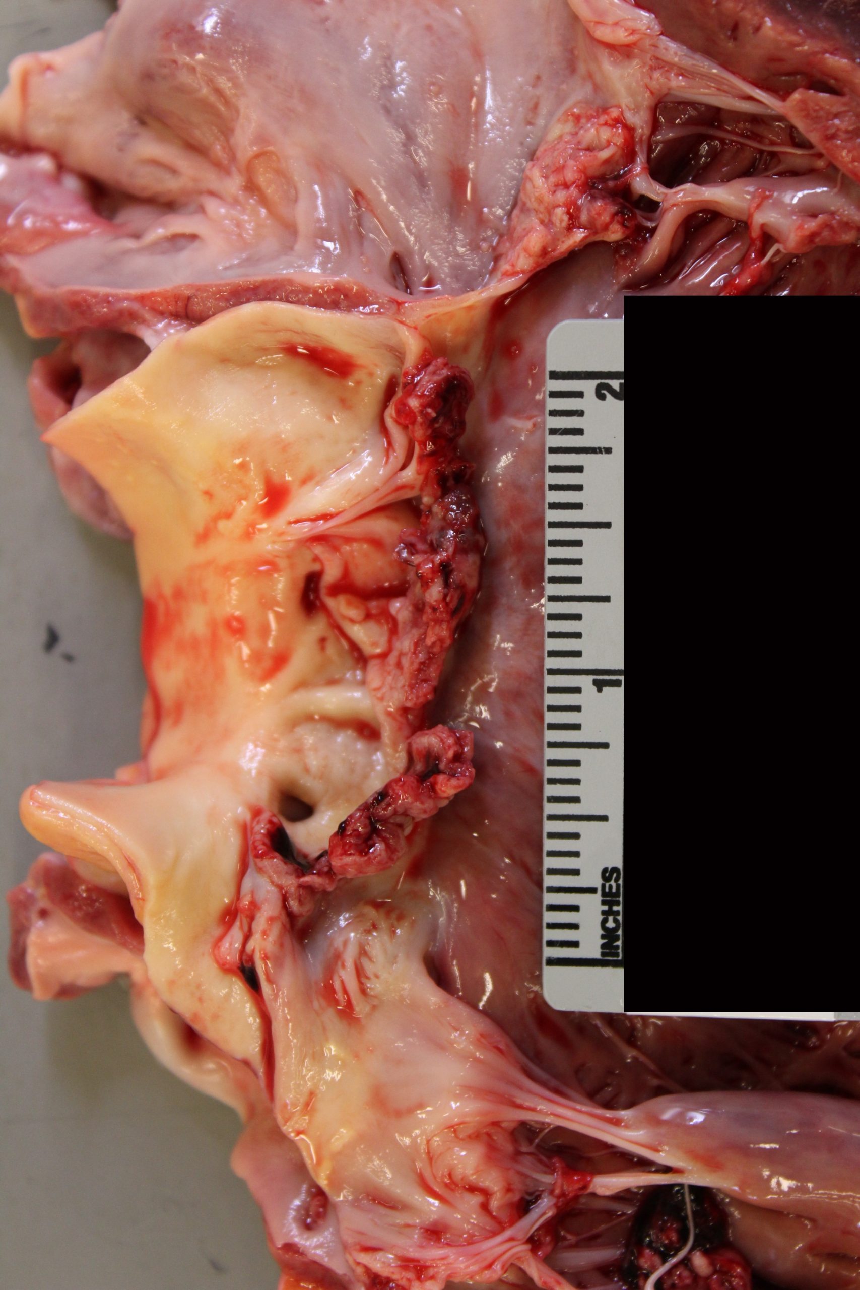
Though they can get pretty large. This one sat over a mechanical valve replacement from a previous vegetation:
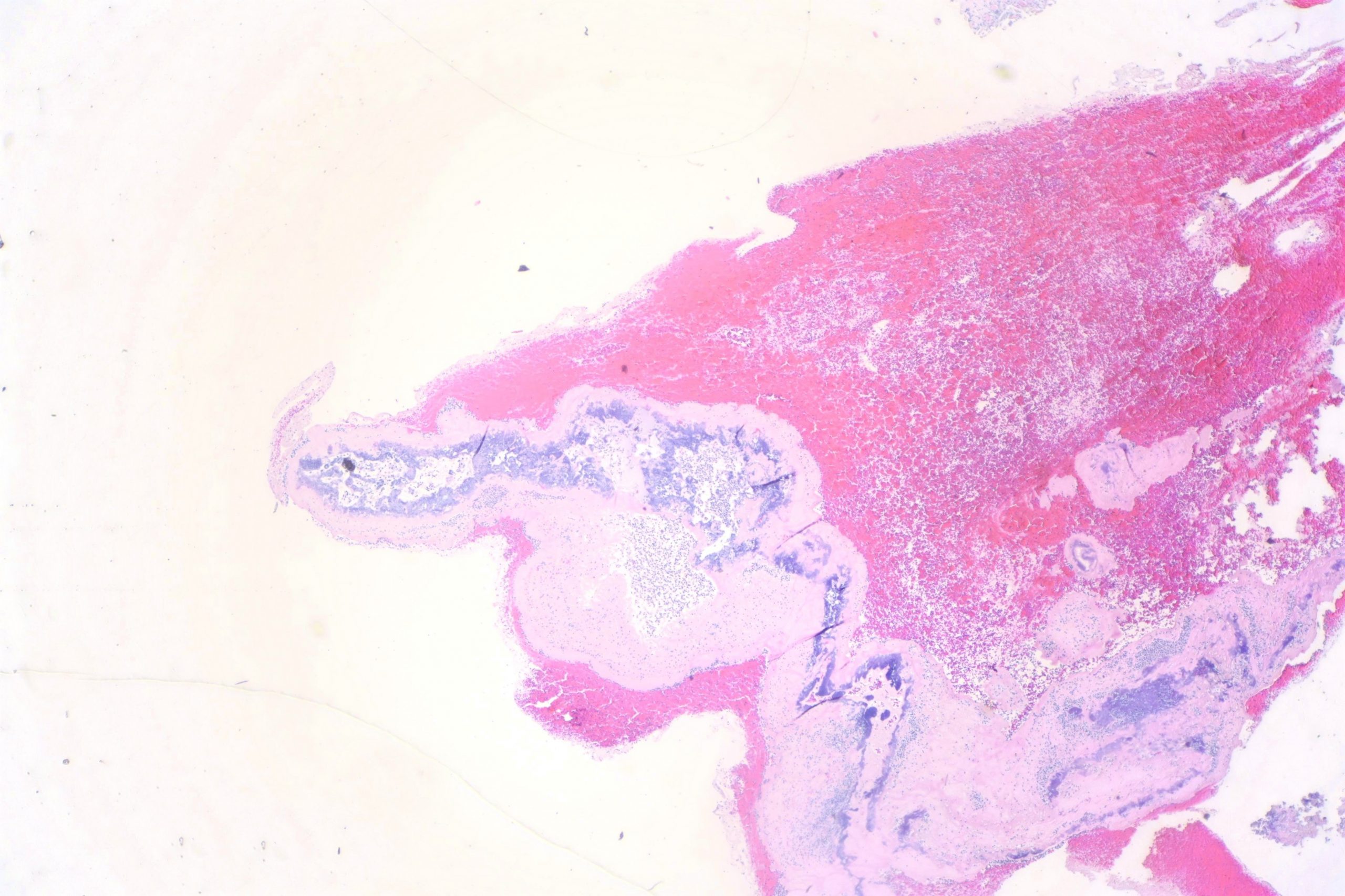
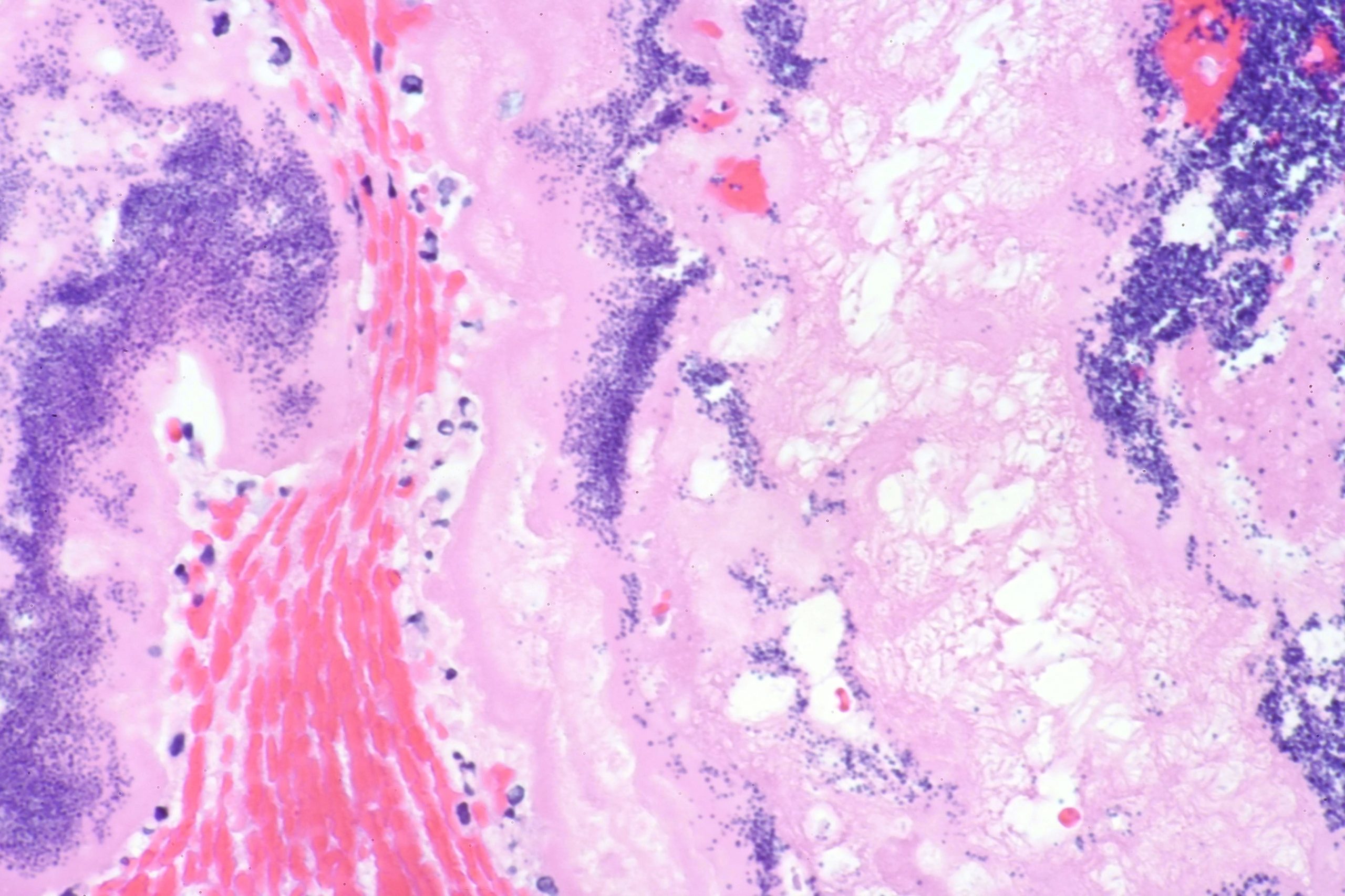
Here’s a typical microscopic appearance of layers of fibrin, inflammatory cells and bacteria:


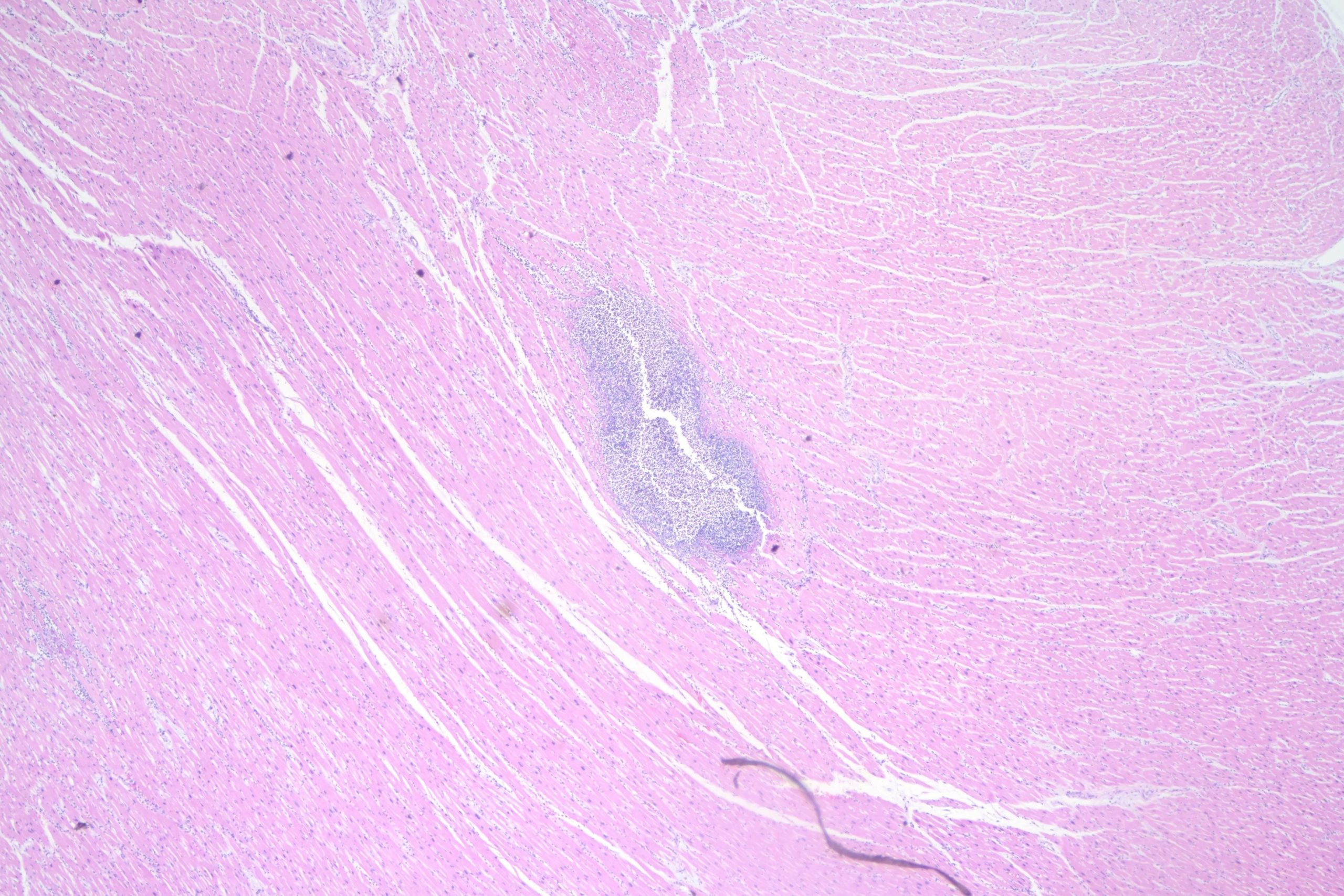
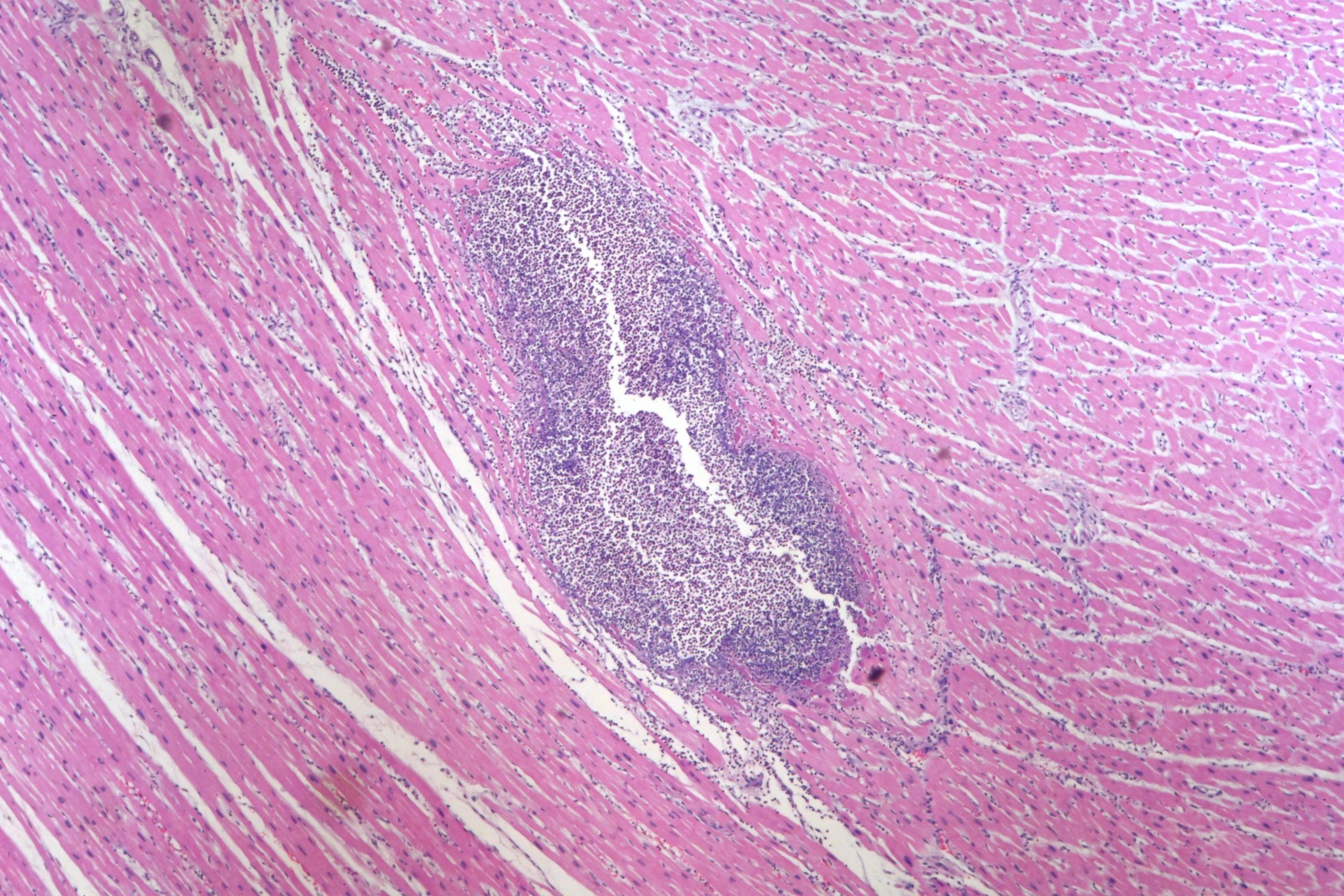
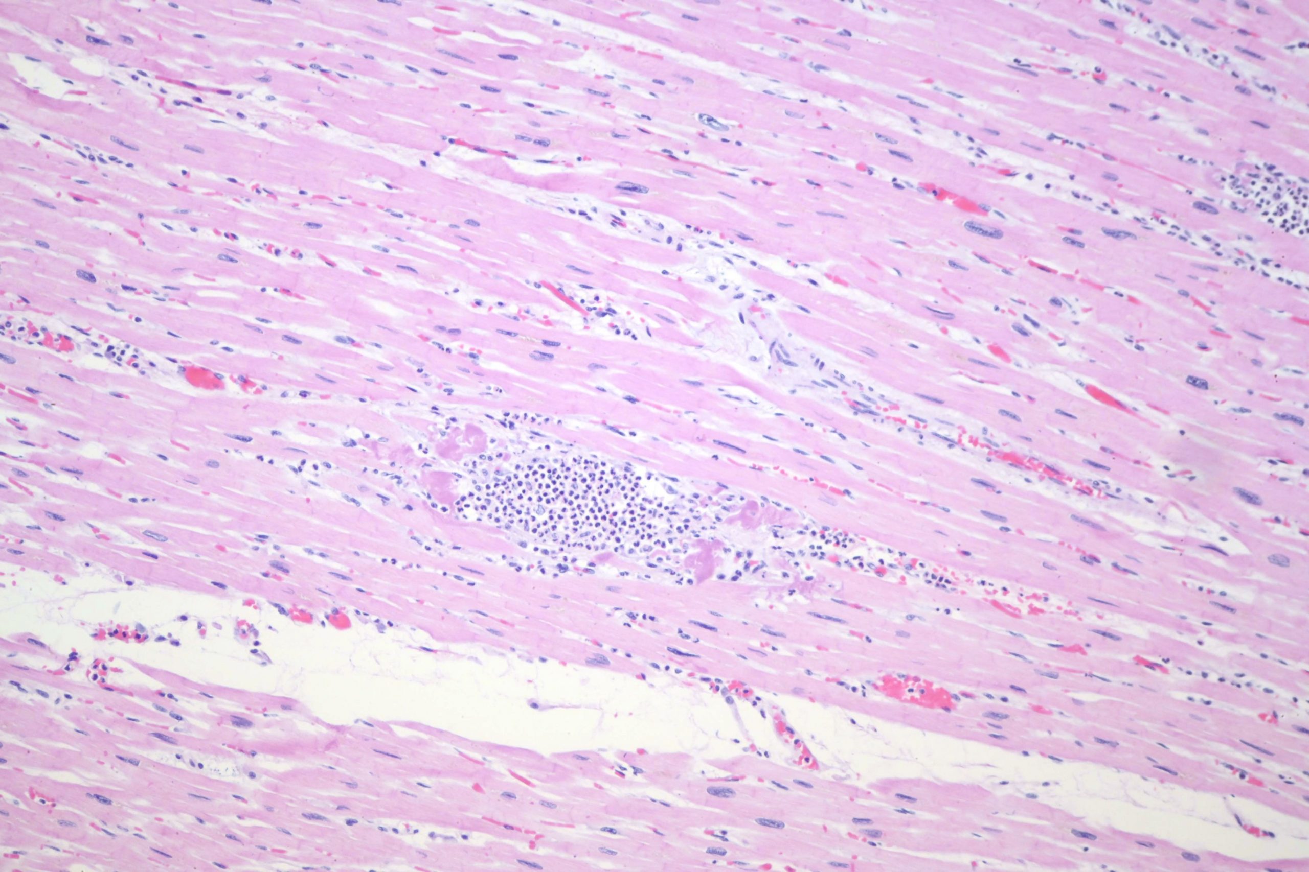
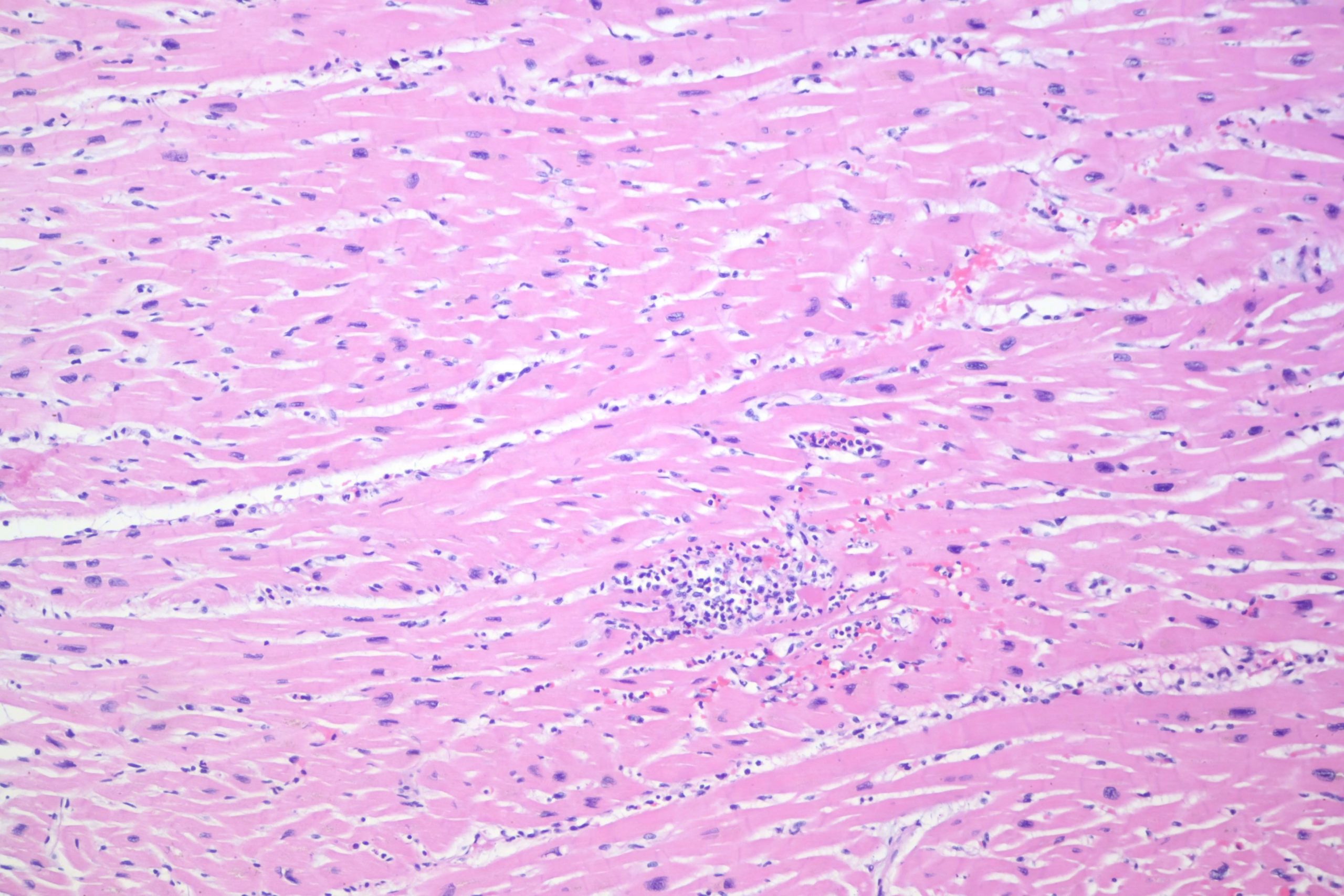
In this case that I made photomics for, there were abscesses in multiple organs, including the heart:
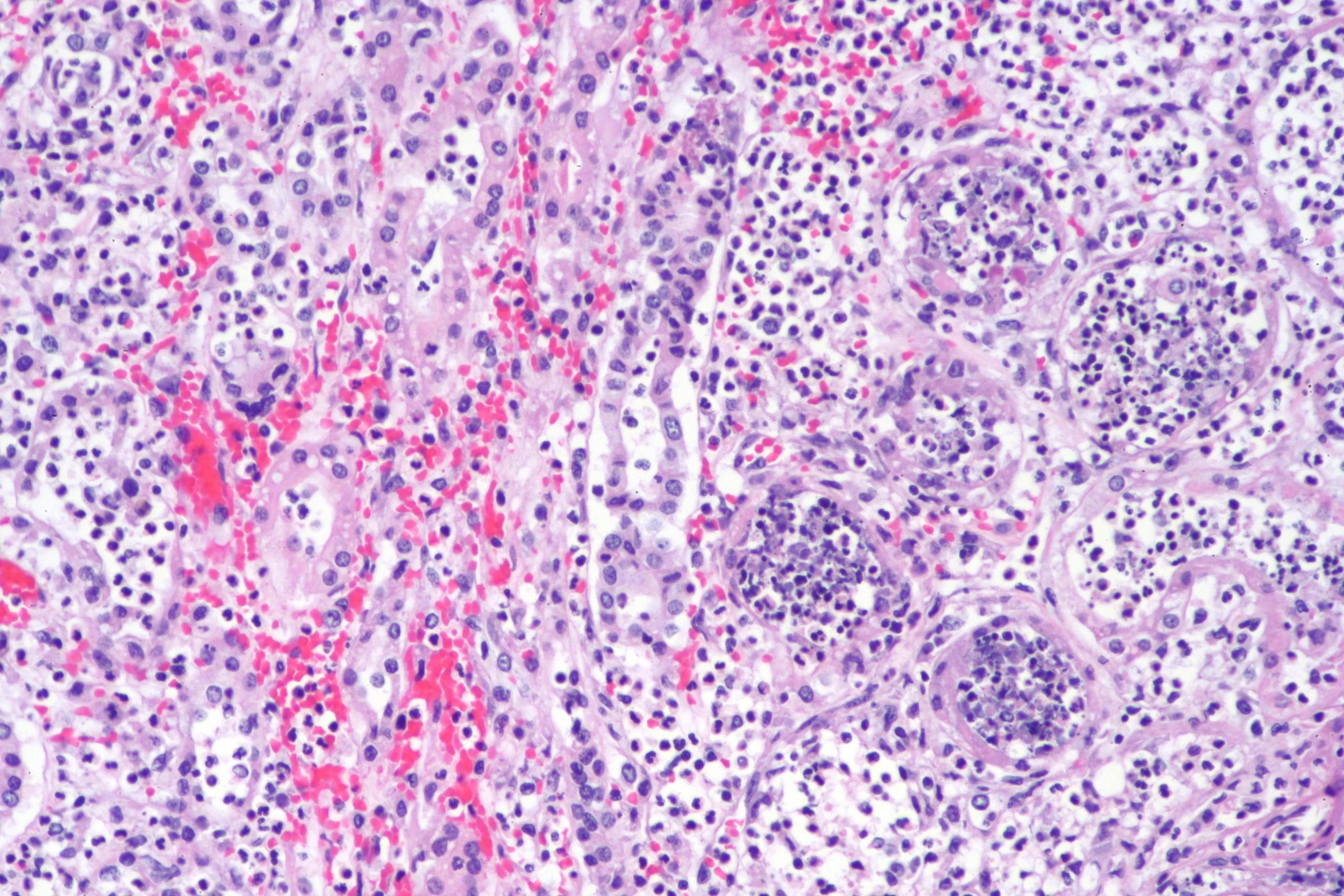
The liver:
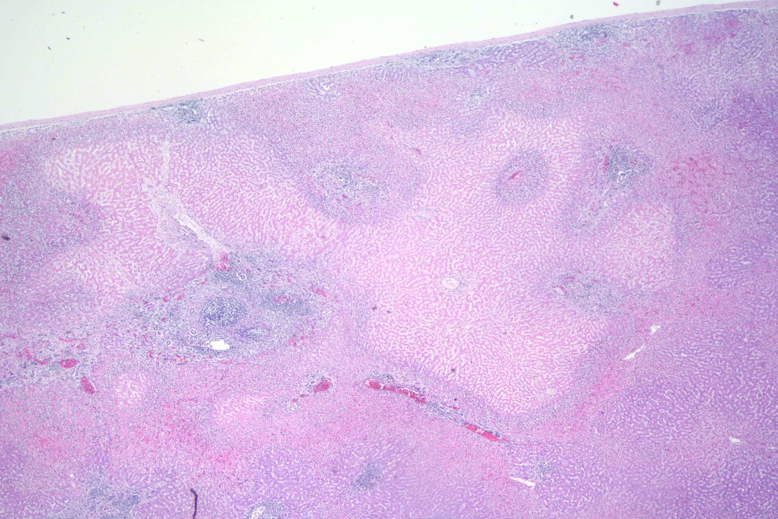
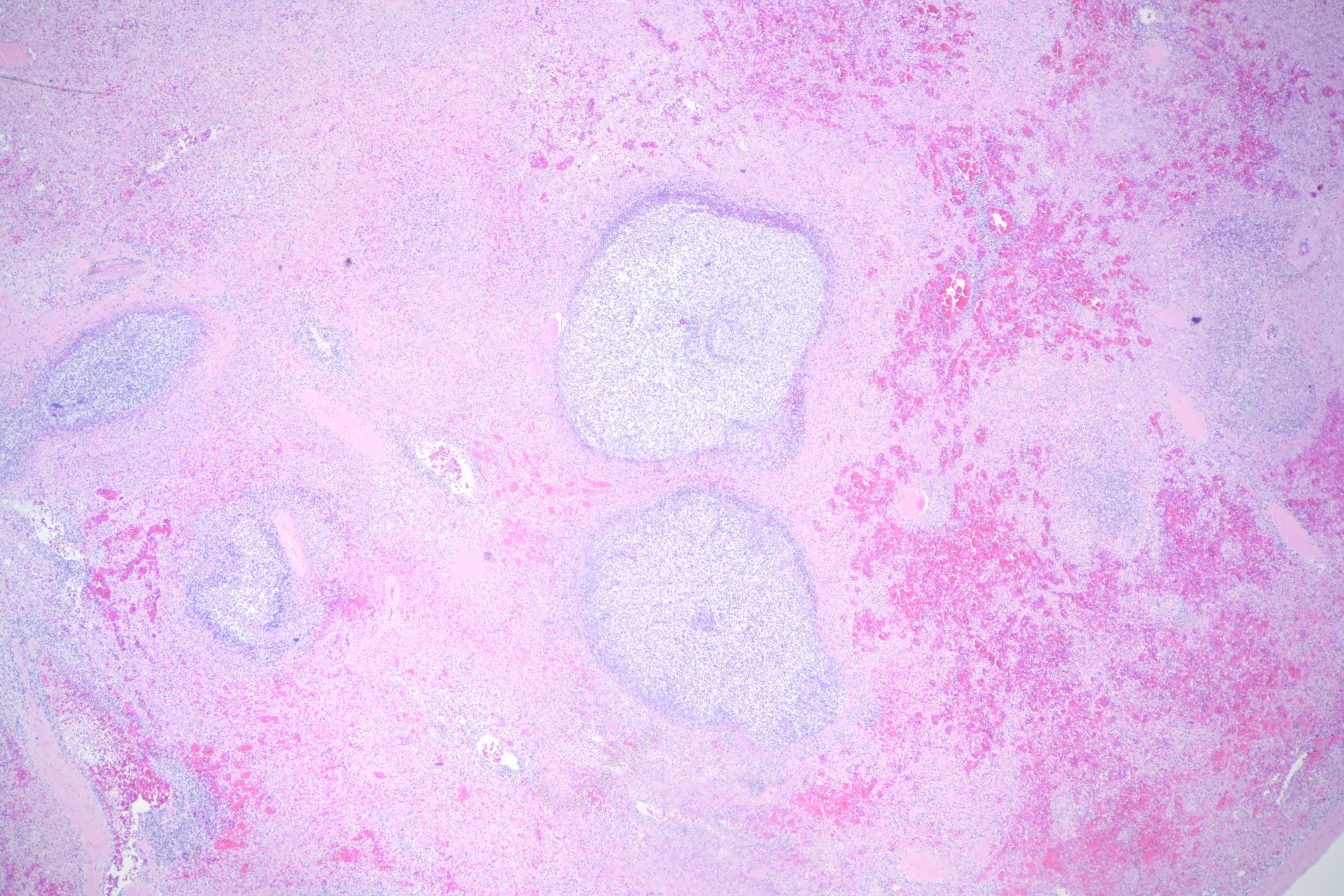
The spleen:
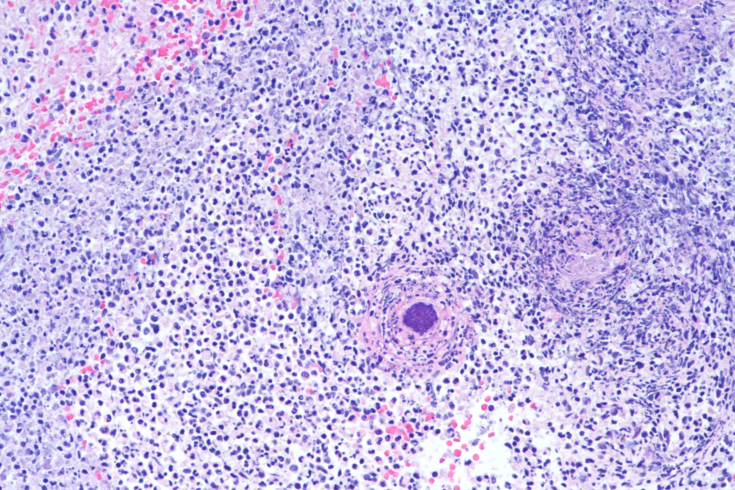
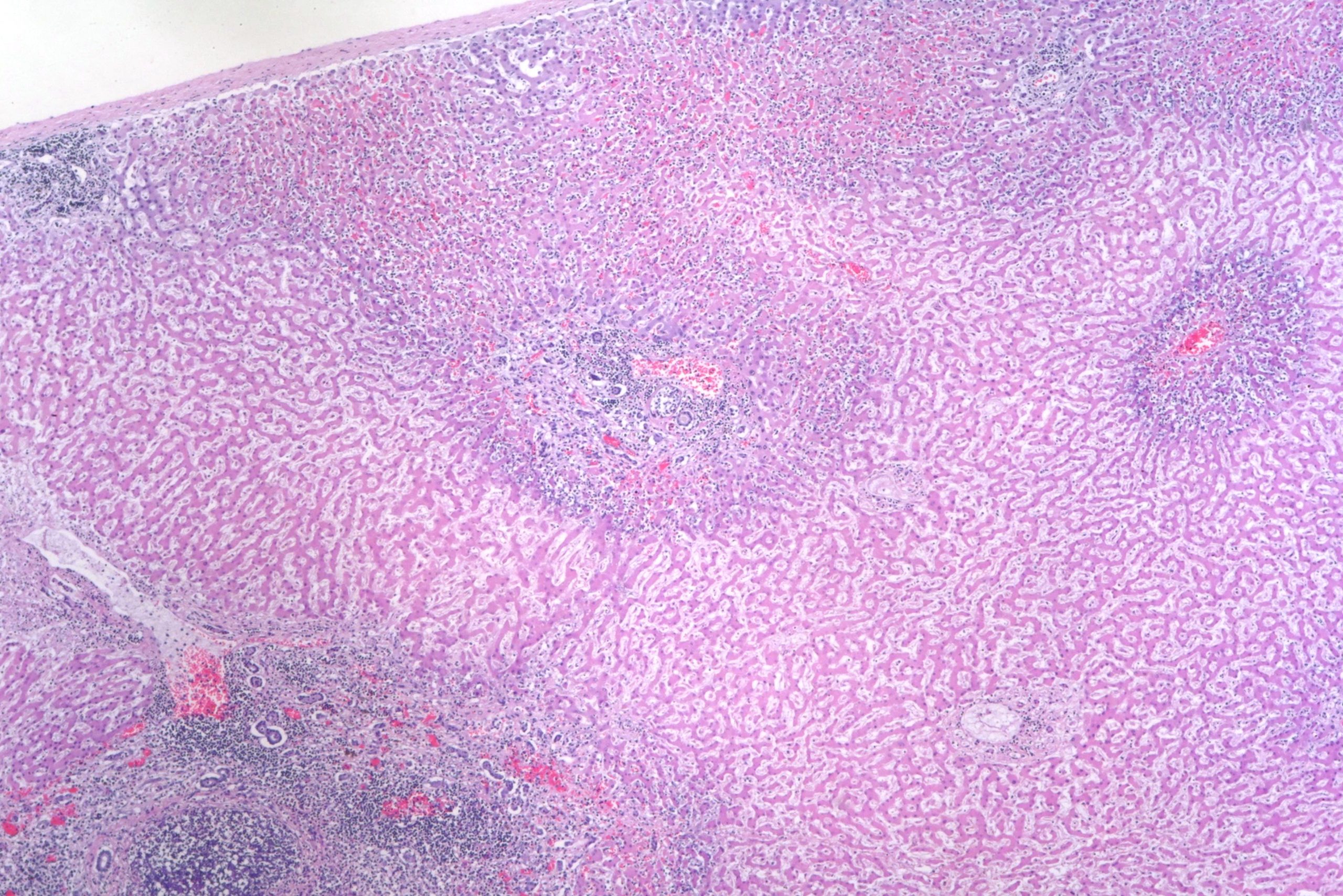
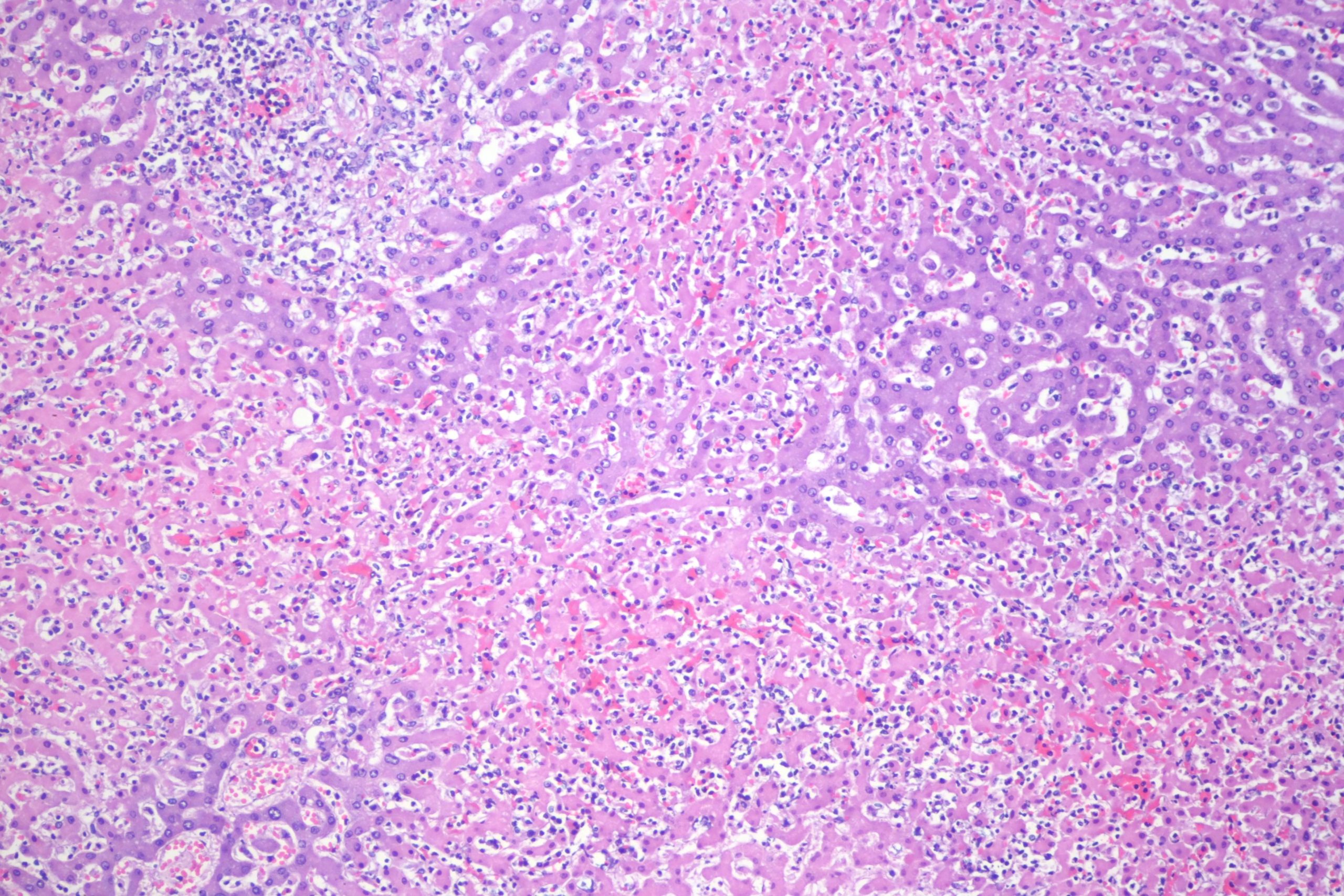
The kidney:


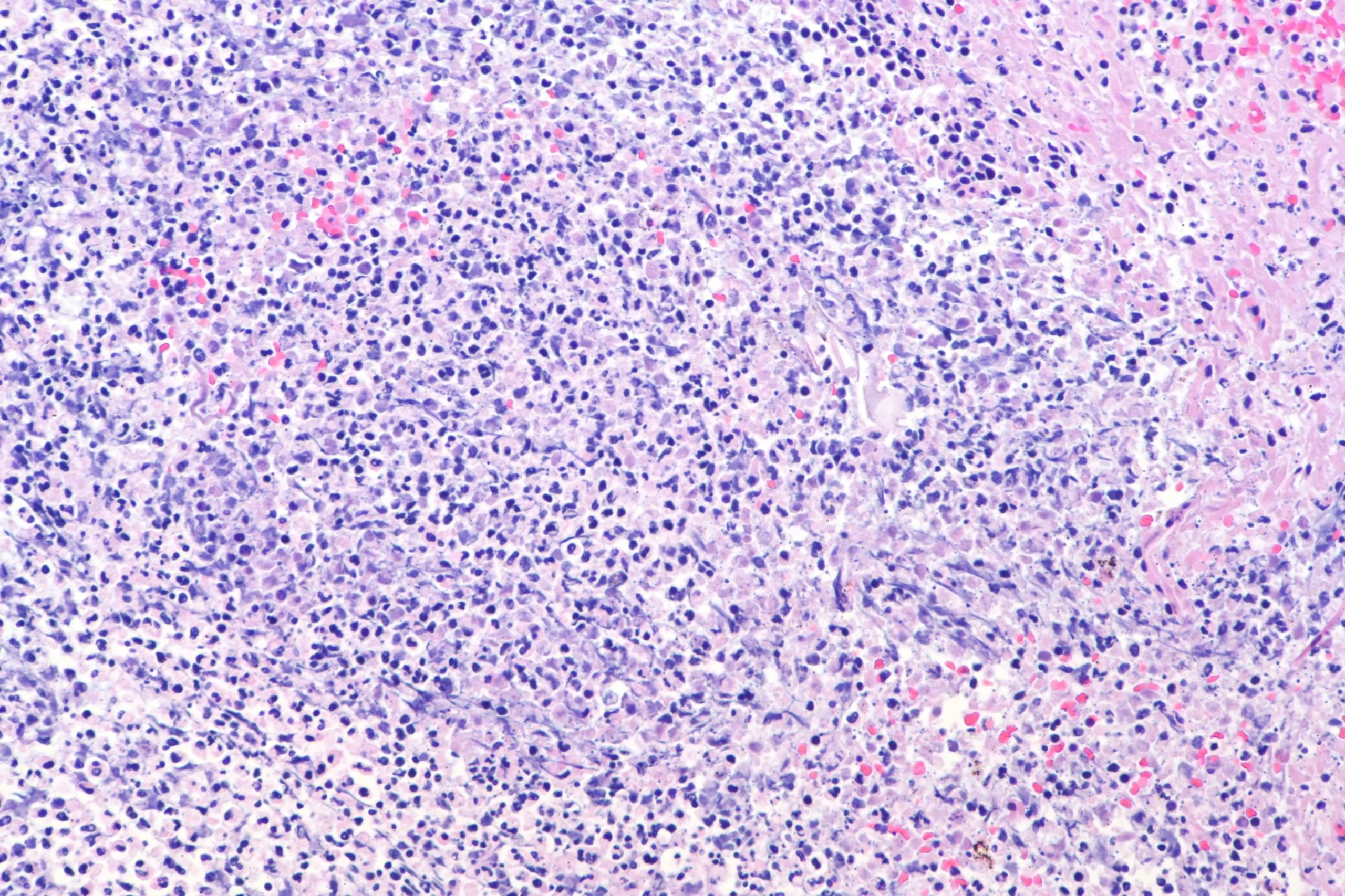
And the brain:

As always, free for use in lecture, or teaching, with or without attribution (though attribution is appreciated). If you put these in a publication, please contact me. Higher resolution images with lossless compression are available on request until I lose them, if you need them for a lecture or such. Email me if you want me to send them to you.
Great for teaching. Thanks!